


|
- Home
- About Us
-
Our Business
-
Medical IVD
- Abbott Bioline CHOLERA Ag 01/0139
- Calbiotech high sensitivity C-Reactive Protein ELISA
- Thermo Fisher Scientific Human RARRES2/TIG2 ELISA Kit
- InBios Kalazar Detect Rapid Test for Visceral Leishmaniasis
- Bioline RSV
- West Nile Detect IgG ELISA
- Abbott Bioline DENGUE IgG/IgM WB
- Abbott Bioline HIV 1/2 3.0
- Boditech ichroma Free T4
- Inbios Kalazar Detect Rapid Test for Visceral Leishmaniasis
- Thermo Fisher Scientific Human EGFR (Full-length) ELISA Kit
- Abbott Bioline HAV IgG/IgM
- Abbott Bioline LEPTOSPIRA IgM
- Abbott Bioline NOROVIRUS
- Abbott Bioline SYPHILIS 3.0
- SD Biosensor ULTRA Malaria P.F/Pan Ag
- Ultra Chikungunya IgM/IgG
- Bioline SALMONELLA TYPHI IgG/IgM FAST
- Abbott Bioline TSUTSUGAMUSHI
- Abbott DETERMINE HBsAg 2
- Abbott Bioline ZIKA IgM
- Abbott Bioline INFLUENZA ULTRA
- Abbott Bioline STREP A
- SD Biosensor Ultra HBsAg
- Abbott Panbio DENGUE IgM CAPTURE ELISA
- Zybio Glutathione Reductase
- Calbiotech Free Testosterone ELISA
- Calbiotech Testosterone Total ELISA
- Abbott Bioline HANTAAN VIRUS
- zybio Apolipoprotein A1
- Abbott DETERMINE HIV -1/2 Ag/Ab Combo
- The West Nile Detect IgM Capture ELISA
- ZIKV Detect 2.0 IgM Capture ELISA Kit
- Boditech ichroma II - Anemia Controls
- ZYBIO Microalbuminuria
- Zybio Prealbumin
- Zybio N-acetyl- -D-glucosaminidase
- Zybio Total Bile Acids
- Zybio Cholyglycine (Homogeneous Enzyme Immune)
- Zybio Cystatin C (Colloidal Gold Immunoturbidimetric)
- Zybio Cholyglycine (Latex Enhanced Immunoturbidimetric)
- Abbott ABON Urine Test E-Z Split Key Drug Screen Cup
- STANDARD M nCoV SD Biosensor Real-Time Detection kit
- ThermoFisher Scientific Accula System Rapid Reliable RT-PCR Test
- Abbott ID NOW Instrument
- InBios Scrub Typhus Detecta c IgG Rapid Test
- Thermo Fisher Taqpath Covid-19 Combo Kit
- Abbott Panbio COVID Antigen Self Test
- Sure Status Covid-19 Antigen Card Test Kit
- iHealth COVID-19 Antigen Rapid Test
- Molbio Truenat COVID 19 Chip based Real Time PCR Test Kit
- STANDARD Q COVID 19 Ag
- Abbott BinaxNOW Covid-19 Rapid Antigen Card Test Kit
- MagMAX Viral Pathogen Nucleic Acid Isolation Kit
- Standard Q Covid - 19 Ag
- TRIVITRON Monkeypox Real Time PCR Test Kit
- RADI Monkeypox Virus Detection Kit
- RADI COVID-19 Detection Kit
- Hi-PCR Coronavirus (COVID-19) Multiplex Probe PCR Kit
- HIPurA Viral RNA Purification Kit
- Ultra Covid - 19 Ag
- Abbott Realtime Sars-Cov-2 Assay
-
Medical RTD
- ABON Syphilis Ultra Rapid Tests (Whole Blood Serum Plasma)
- Erba Mannheim ErbaQik HCV Test Card
- Erba Mannheim ErbaQik Sickle Cell Test Card
- CTK Biotech OnSite Toxo IgG/IgM Combo Rapid Test
- CTK Biotech OnSite S. Typhi/Paratyphi Ag Rapid Test
- Abbott Bioline CHLAMYDIA
- CTK Biotech OnSite Typhoid IgG/IgM Rapid Test
- CTK Biotech Onsite Typhoid IgG/IgM Combo Rapid Test CE
- SD Biosensor STANDARD Q TSUTSUGAMUSHI IgM/IgG
- Roche Roche cardiac Troponin T sensitive test (visual)
- Abbott DETERMINE HIV-1/2 Set
- Acro Biotech JusChek Multi-Drug 6 Drugs Rapid Test Cassette (Urine)
- CTK Biotech OnSite TORCH Panel Rapid Test
- CTK Biotech OnSite Syphilis Ab Rapid Test (strip) CE
- Abbott Bioline ROTA/ADENO
- Abbott DETERMINE SYPHILIS TP
- Abbott BINAXNOW STREP A
- Abbott Bioline RUBELLA IgG/IgM
- Abbott DETERMINE TB LAM Ag
- Abbott SURESTEP URINE DRUG TEST E-Z SPLIT KEY CUP
- CTK Biotech TRUSTline Scrub typhus IgG/IgM Rapid Test
- Acro Biotech JusChek COT200 Rapid Test Cassette (Urine)
- Thermo Fisher Scientific Thermo Scientific SickleScreen Assay Kit (120T)
- Thermo Fisher Scientific Thermo Scientific SickleScreen Assay Kit (1T)
- Acro Biotech JusChek COT10 Rapid Test Cassette (Whole Blood/ Serum/ Plasma)
- HemoTypeSC HemoTypeSC Sickle Cell Disease
- Acro Biotech JusChek Alcohol (ALC) Rapid Test Cassette
- SD Biosensor STANDARD Q Malaria P.F/Pan Ag
- Acon ACON hCG (Urine)
- SD Biosensor STANDARD Q HIV 1/2 Ab
- CTK Biotech OnSite H. pylori Ab Combo Rapid Test
- Abbott Bioline MALARIA Ag P.f/Pan
- Abbott Bioline INFLUENZA Ag A/B/A (H1N1)
- CTK Biotech OnSite Leptospira IgG/IgM Combo Rapid TestTest
- SD Biosensor STANDARD Q Ultra-Dot HIV
- SD Biosensor STANDARD Q Malaria P.f Ag
- OraSure Technologies ORAQUICK HCV RAPID ANTIBODY TEST
- Abbott Bioline LEPTOSPIRA IgG/IgM
- Premier Medical First Response Malaria Ag pf/pv Card Test
- Abbott Bioline H. PYLORI Ag
- Premier Medical FIRST RESPONSE HIV 1-2.O CARD TEST (SELF TEST)
- CTK Biotech OnSite HAV IgG/IgM Rapid Test CE
- Abbott PANBIO HIV SELF TEST
- Abbott Bioline HIV/SYPHILIS DUO
- SD Biosensor STANDARD Q Ultra Dot HCV
- SD Biosensor STANDARD Q HBsAg
- Abbott BINAXNOW LEGIONELLA URINARY ANTIGEN CARD
- CTK Biotech OnSite H. pylori Ag Rapid Test
- CTK Biotech OnSite Dengue IgG/IgM 3.0 Combo Rapid Test CE
- InBios Dengue NS1 Detect Rapid Test
- Abbott Bioline EV71 IgM
- CTK Biotech OnSite Dengue Ag Rapid Test CE
- SD Biosensor STANDARD Q Dengue IgM/IgG
- Abbott Bioline DENGUE NS1 Ag
- SD Biosensor STANDARD Q COVID/Flu Ag Combo
- Abbott BinaxNOW G6PD
- SD Biosensor STANDARD Q Dengue Ag+Ab Duo
- SD Biosensor Ultra Covid-19 Ag
- SD Biosensor STANDARD Q COVID-19 IgM/IgG Combo
- SD Biosensor STANDARD Q COVID-19 Ag
- SD Biosensor STANDARD Q Filariasis Ag
- InBios CL Detect Rapid Test for Cutaneous Leishmaniasis
- CTK Biotech TRUSTline Filariasis IgG/IgM Rapid Test
- Premier Medical Sure Status Covid-19 Antigen Card Test Kit
- Roche COVID-19 At Home Test
- Abbott Panbio COVID-19 Antigen Self Test
- SD Biosensor STANDARD i-Q COVID/Flu Ag Combo Test
- Abbott DETERMINE Chase Buffer
- InBios Chagas Detect Plus Rapid Test
- Acon Flowflex SARS-CoV-2 Antigen Rapid Test
- Abbott PANBIO COVID-19/FLU A&B RAPID PANEL (NASOPHARYNGEAL)
- CTK Biotech OnSite Chikungunya IgM Combo Rapid Test
- SD Biosensor STANDARD Q Malaria P.F/P.V Ag
- Erba Mannheim ErbaQik Malaria Pan Antigen test (Pan pLDH)
- Abbott Bioline MALARIA Ag P.f/P.f/P.v
- Abbott BINAXNOW MALARIA
- Abbott Bioline LEGIONELLA Ag
- Abbott Bioline ONCHO/LF IgG4 BIPLEX
- Abbott DETERMINE HIV EARLY DETECT SET
- Abbott Bioline LYMPHATIC FILARIASIS IgG4
- Abbott Bioline ROTAVIRUS
- Premier Medical First Response Malaria Ag pLDH/HRP2 Combo Card Test
- Acro Biotech JusChek Alcohol (ALC) Rapid Test Dipstick
- Abbott CheckNOW HIV SELF TEST
- SD Biosensor STANDARD Q Chikungunya IgM/IgG
- Abbott Determine HBsAg 2 SET
- Abbott Bioline LEISHMANIA Ab
- SD Biosensor STANDARD Q Leptospira IgM/IgG
- CTK Biotech OnSite RF Rapid Test
- CTK Biotech OnSite FOB-Hi Rapid Test
- First Response Premier MedicalSyphilis Anti TP Card Test
- OraSure Technologies ORAQUICK IN-HOME HIV TEST
- Abbott DETERMINE HIV EARLY DETECT
- SD Biosensor STANDARD Q HIV/Syphilis Combo
- Abbott Bioline MALARIA Ag P.f/P.v
- Premier Medical First Response HIV 1+2/Syphilis Combo Card Test
- Abbott ABON COT One Step Cotinine Test Device Urine
- Acro Biotech JusChek Multi - Drug 10 Drugs Rapid Test Cassette (Urine)
- CTK Biotech OnSite Troponin I Combo Rapid Test
- CTK Biotech OnSite HAV IgM Rapid Test
- Abbott DETERMINE HBsAg 2
- Acro Biotech JusChek Alcohol (ALC) Rapid Test Dipstick (Saliva)
- Erba Mannheim ErbaQik HBsAg Test Card
- Erba Mannheim ErbaQik Dengue IgG/IgM
- Erba Mannheim ErbaQik Malaria Pf/Pv Ag (HRP2/Pv pLDH)
- Erba Mannheim ErbaQik Syphilis Test Card
- Abbott DETERMINE SYPHILIS TP SET
- Erba Mannheim ErbaQik Malaria Pf/Pan Ag (HRP2/Pan pLDH)
- FIRST RESPONSE HIV 1 - 2.0 CARD TEST
- Abbott Bioline Filariasis Test Strip
- ABON hCG One Step Pregnancy Tests - For Professional Use
- Bioline DENGUE NS1 Ag
- Bioline TB Ag MPT64
- FIRST RESPONSE Malaria Ag. pLDH HRP2 Combo Card Test
- Premier Medical FIRST RESPONSE Malaria Antigen P. faciparum(HRP2) Card Test
- Roche Cardiac Trop T Sensitive Test
- First Response SyphiliS Anti-TP CardTest
- FIRST RESPONSE HIV 12 SYPHILIS Combo Card Test
- Bioline Dengue Duo
- First Response Malaria Ag. P.f. P.v. Card Test
- Binaxnow Streptococcus Pneumoniae
- Bioline HCV
- Determine HIV-1 2
- BinaxNOW LEGIONELLA URINARY ANTIGEN CARD
- Determine HBsAg
- Bioline HIV 1 2 3.0
- FIRST RESPONSE Malaria Antigen P. faciparum(HRP2) Card Test
- Bioline Malaria Ag P.f Pan
- Bioline Malaria Ag P.f Pv
- ACON hCG (Urine)
- Determine Combo 4th Generation Test Kit For Clinical
- Bioline DENGUE IgM CAPTURE ELISA
- iScript Reverse Transcription Supermix
- Abbott BINAXNOW STREPTOCOCCUS PNEUMONIAE ANTIGEN CARD
- Erba Mannheim ErbaQik Dengue Duo
- Abbott Bioline HAT 2.0
- Abbott Bioline HBsAg
- SD Biosensor STANDARD Q RSV Ag
- Abbott Bioline INFLUENZA ANTIGEN
- Erba Mannheim ErbaQik Dengue NS1 Ag
- Abbott Bioline H. PYLORI Ab
- SD Biosensor STANDARD Q HCV Ab
- Abbott Bioline HCV
- SD Biosensor STANDARD Q Influenza A/B
- Acro Biotech JusChek COT20 Rapid Test (Oral Fluid)
- CTK Biotech Onsite HEV IgM Rapid Test CE
- Roche SARS-CoV-2 Rapid Antigen Test.
- SD Biosensor STANDARD Q Dengue NS1 Ag
- Abbott BINAXNOW COVID-19 Ag CARD
- Abbott NMP22 BLADDERCHEK
- Premier Medical First Response Syphilis Anti TP Card Test
- Abbott Panbio COVID-19 Ag Rapid Test Device (Nasopharyngeal)
- Abbott DETERMINE HIV-1/2
- OnSite Duo Dengue Ag IgG IgM Rapid Test
- STANDARD Q Tsutsugmushi IgM IgG
- STANDARD Q Malaria P.f Pan Ag
- STANDARD Q Dengue Ag Ab Duo
- STANDARD Q Malaria PF PV Ag
- Standard Q Filariasis Ag
- STANDARD Q Dengue IgM IgG
- OnSite FOB-Hi Rapid Test
- OnSite HEV IgM Rapid Test
- OnSite H.Pylori Ag Rapid Test
- OnSite HAV IgM Rapid Test
- OnSite HAV IgG IgM Rapid Test
- OnSite Leptospira IgG IgM Combo Rapid Test
- TRUST line Filariasis IgG IgM Combo Rapid Test
- OnSite S.Typhi Paratyphi Ag Rapid Test
- OnSite Typhoid IgG IgM Rapid Test
- OnSite Dengue IgG IgM Combo Rapid Test
- OnSite Dengue Ag Rapid Test
- STANDARD Q HIV 1-2 Ab Test
- STANDARD Q Chikungunya IgM IgG
- STANDARD Q Ultra-Dot HCV
- STANDARD Q Hbsag
- STANDARD Q Ultra-Dot HIV
- STANDARD Q HCV Ab
- STANDARD Q Dengue NS1 Ag
- OnSite H. Pylori Ab combo Rapid Test
- STANDARD Q - HIV Syphilis Combo
- OnSite Toxo IgG IgM Combo Rapid Test
- Standard Q Lepospira Igg Igm
- N SURE One step Pragnancy Detention Test
- OnSite RF Rapid Test
- OnSite TORCH Panel Rapid Test
- Dengue Day 1 Test
- Advantage MAL Card
- CTK TRUSTline Scrub typhus IgG IgM Rapid Test
- HCV TRI DOT
- HIV TRI DOT
- Hepa card
- Bioline CHAGAS Ab
- PANBIO COVID-19 IgG/IgM RAPID TEST DEVICE
- STANDARD Q COVID-19 Ag
- CTK Biotech OnSite Duo Dengue Ag-IgG/IgM Rapid Test CE
- Abbott PANBIO HIV VERIFICATION TEST
- ORAQUICK IN-HOME HIV TEST (Community Version)
- ORAQUICK IN-HOME HIV TEST (Pharmacy Version)
- ORAQUICK IN-HOME HIV TEST (US FDA Version)
- Abbott BINAXNOW INFLUENZA A&B CARD
- Abbott Bioline MALARIA Ag P.f (HRP2/pLDH)
- Abbott NxTek ELIMINATE MALARIA Pf
- Abbott BINAXNOW RSV CARD
- Abbott TESTPACK PLUS WITH OBC STREP A
- Erba Mannheim ErbaQik HIV 1+2 Test Card
-
Medical ELISA
- Calbiotech Thyroxine (T4) ELISA
- Calbiotech AMH ELISA
- Calbiotech Toxoplasma IgG ELISA
- Chlamydia trachomatis IgG ELISA
- Chlamydia trachomatis IgM ELISA
- Calbiotech SSB IgG ELISA
- SD Biosensor STANDARD E TB-Feron Tubes 300
- CTK Biotech RecombiLISA Leptospira IgM ELISA
- Abbott Bioline DENGUE NS1 Ag ELISA
- Calbiotech Beta2 Microglobulin ELISA
- SD Biosensor STANDARD E Dengue IgM Capture ELISA
- Calbiotech Dengue Virus IgM ELISA
- Calbiotech Sm IgG ELISA
- Calbiotech Cardiolipin IgG ELISA
- Calbiotech Neonatal TSH ELISA
- CTK Biotech TRUSTwell Free T4 (fT4) ELISA kit
- Calbiotech Insulin ELISA
- Calbiotech C-Peptide ELISA
- Calbiotech Troponin I ELISA
- Calbiotech VZV IgM ELISA
- Calbiotech Rubella IgG ELISA
- Calbiotech Luteinizing Hormone (LH) ELISA
- Calbiotech HSV-1 IgM ELISA
- Calbiotech Neonatal 17(OH) Progesterone ELISA
- Calbiotech Thyroglobulin Ab (TG) ELISA
- Calbiotech Cardiolipin IgG, IgM, IgA ELISA
- Calbiotech Thyroid Peroxidase (TPO) IgG ELISA
- Calbiotech hCG ELISA
- Calbiotech Brucella IgG ELISA
- Calbiotech SSA IgG ELISA
- Calbiotech Mitochondrial (MA) Ab ELISA
- CTK Biotech TRUSTwell Vitamin-D (Total) ELISA
- Calbiotech Cardiolipin IgM ELISA
- Calbiotech HSV-1 IgG ELISA
- Calbiotech CMV IgM ELISA
- InBios Scrub Typhus Detect IgM ELISA Kit
- Calbiotech HE4 ELISA
- SD Standard E Dengue IgG Elisa Analyzes Test Kit
- CTK Biotech TRUSTwell Ferritin ELISA
- Standard E Dengue Elisa Analyzes IgM Test Kit
- Standard E Dengue NS1 Elisa Analyzes Test Kit
- Calbiotech CEA ELISA
- InBios DENV Detect NS1 ELISA
- Chlamydia trachomatis IgA ELISA
- Calbiotech Progesterone ELISA
- CTK Biotech RecombiLISA HAV IgM ELISA CE
- CTK Biotech RecombiLISA HEV IgM ELISA CE
- Calbiotech HSV-2 IgG ELISA
- Calbiotech HSV-2 IgM ELISA
- Calbiotech Cortisol ELISA
- Calbiotech Toxoplasma IgM ELISA
- Calbiotech Cardiolipin IgA ELISA
- Erba Mannheim System Pack LDL-Cholesterol with Calibrator
- Scrub Typhus Detect IgM ELISA Kit
- Calbiotech Free Triiodothyronine (FT3) ELISA
- Calbiotech PSA ELISA
- Calbiotech Ferritin ELISA
- Calbiotech Folate ELISA
- Calbiotech CA 19-9 ELISA
- Calbiotech Brucella IgM ELISA
- RecombiLISA Leptospira IgM ELISA Kit
- Calbiotech AFP ELISA
- Calbiotech Vitamin B12 ELISA
- Calbiotech Scl-70 ELISA
- Calbiotech VZV IgG ELISA
- Calbiotech Intact PTH (Parathyroid Hormone) ELISA
- Calbiotech Immunoglobulin E (IgE) ELISA
- Calbiotech dsDNA IgG ELISA
- Calbiotech 25(OH) Vitamin D ELISA
- Calbiotech CA 15-3 ELISA
- Calbiotech CA-125 ELISA
- Calbiotech Free Beta hCG ELISA
- Calbiotech CMV IgG ELISA
- Calbiotech Bio Intact PTH ELISA
- Calbiotech Free PSA ELISA
- Calbiotech DHEA-S ELISA
- Calbiotech RNP/Sm Autoantibody ELISA
- Calbiotech Cotinine ELISA
- Calbiotech ANA Screen IgG ELISA
- RecombiLISA Dengue Ag ELISA Kit
- TRUSTwell CHIK IgM ELISA Kit
- TRUSTwell HBsAg ELISA Kit
- TRUSTwell HIV Ag-Ab ELISA Kit
- TRUSTwell HIV 1 2 Ab ELISA Kit
- TRUSTwell Dengue IgG ELISA Kit
- Scrub Typhus Detect IgG ELISA System
- CHIKjj Detect IgG ELISA Kit
- Filaria Detect IgG4 ELISA
- ZIKV Detect 2.0 IgM Capture ELISA
- RecombiLISA HAV IgM ELISA Kit
- RecombiLISA Total T3 Elisa Kit
- RecombiLISA Total T4 ELISA Kit
- RecombiLISA TSH ELISA kit
- TRUSTwell Dengue IgM ELISA Kit
- TRUSTwell Syphilis Ab kit
- TRUSTwell HCV IgG ELISA Kit
- STANDARD E TB-Feron Tubes 200
- STANDARD E TB-Feron Tubes 300
- J Mitra ELISA Reader
- J Mitra ELISA Washer
- DENV Detect NS1 ELISA
- InBios DENV Detect IgM Capture ELISA
- CHIKjj Detect IgM ELISA Kit
- RecombiLISA HEV IgM ELISA Kit
- CTK Biotech TRUSTwell CHIK IgM ELISA CE
- Chlamydia pneumonia IgG ELISA
- Chlamydia pneumonia IgM ELISA
- CTK Biotech TRUSTwell COVID-19 IgG ELISA
- Abbott Bioline DENGUE IgG CAPTURE ELISA
- Abbott BIOLINE DENGUE IgM CAPTURE ELISA
- Abbott Panbio DENGUE IgG INDIRECT ELISA
- Calbiotech Dengue Virus IgG ELISA
- CTK Biotech TRUSTwell Dengue Ag ELISA
- CTK Biotech TRUSTwell Dengue IgG ELISA CE
- CTK Biotech TRUSTwell Dengue IgM ELISA CE
- SD Biosensor STANDARD E Dengue IgG Capture ELISA
- SD Biosensor STANDARD E Dengue NS1 Ag ELISA
- Calbiotech EBV-VCA IgG ELISA
- Calbiotech EBV-VCA IgM ELISA
- Calbiotech Follicle Stimulating Hormone (FSH) ELISA
- Calbiotech Prolactin ELISA
- CTK Biotech TRUSTwell Anti-Mullerian hormone (AMH) ELISA
- Calbiotech 17(OH) Progesterone ELISA
- Calbiotech Androstenedione ELISA
- Calbiotech Estradiol ELISA
- Calbiotech Growth Hormone ELISA
- Calbiotech H. Pylori IgA ELISA
- Calbiotech H. Pylori IgG ELISA
- Calbiotech H. Pylori IgM ELISA
- CTK Biotech TRUSTwell HBsAg ELISA
- CTK Biotech TRUSTwell HCV Total Ab ELISA
- Athenese-Dx TRUSTwell HIV 1+2 Ab ELISA
- CTK Biotech TRUSTwell HIV Ag-Ab ELISA
- Calbiotech C-Reactive Protein ELISA
- Abbott Panbio LEPTOSPIRA IgM ELISA
- CTK Biotech TRUSTwell Malaria Ag ELISA
- Calbiotech Measles IgG ELISA
- Calbiotech Measles IgM ELISA
- Calbiotech Mumps IgG ELISA
- Calbiotech Mumps IgM ELISA
- Calbiotech Mycoplasma IgG ELISA
- Calbiotech Mycoplasma IgM ELISA
- Calbiotech Neonatal Galactose ELISA
- Calbiotech Neonatal Maple Syrup Urine Disease (N-MSUD) ELISA
- Calbiotech Neonatal PKU ELISA
- Abbott Bioline ONCHOCERCIASIS IgG4 ELISA
- Calbiotech Rubella IgM ELISA
- Calbiotech Salmonella IgG ELISA
- Calbiotech Salmonella IgM ELISA
- Calbiotech Treponima pallidum IgG ELISA
- CTK Biotech TRUSTwell Syphilis Ab ELISA CE
- SD Biosensor STANDARD E TB-Feron Tubes 200
- Calbiotech Free Thryoxine (FT4) ELISA
- Calbiotech Triiodothyronine (T3) ELISA
- Calbiotech TSH ELISA
- CTK Biotech TRUSTwell Free T3 ELISA Kit
- CTK Biotech TRUSTwell Total T3 Elisa Kit
- CTK Biotech TRUSTwell Total T4 ELISA Kit
- CTK Biotech TRUSTwell TSH ELISA Kit
- Qiagen QuantiFERON-TB Gold Plus Blood Collection Tubes
- InBios CHIKjj Detect IgG ELISA Kit
- InBios CHIKjj Detect IgM ELISA Kit
- InBios DENV Detect IgG ELISA
- InBios DENV Detect IgM Capture ELISA Kit
- Inbios Filaria Detect IgG4 ELISA
- InBios JE Detect IgG ELISA
- InBios JE Detect IgM Antibody Capture ELISA
- InBios Scrub Typhus Detect IgG ELISA Kit
- InBios West Nile Detect IgG ELISA
- InBios West Nile Detect IgM Capture ELISA
- InBios ZIKV Detect 2.0 IgM Capture ELISA Kit
-
Medical FIA
- ichroma Rheumatoid Arthritis (RF IgM)
- ichroma Human Chorionic Gonadotropin
- Finecare Testosterone Rapid Quantitative Test
- ichroma ST2
- Getein Novel Coronavirus (2019-nCoV) IgM/IgG Antibody Fast Test Kit
- ichroma D Dimer Test
- Standard F Dengue NS1 Ag FIA
- Finecare Fia Meter lll Plus
- wondfo Finecare CK-MB Rapid Quantitative Test
- ichroma Interleukin-6 (IL-6)
- Getein 1100 Immunofluorescence Quantitative Analyzer
- Wondfo Finecare FIA Meter III Plus
- STANDARD F H. Pylori Ag FIA
- ichroma Anti-CCP Plus
- ichroma Creatine Kinase Muscle Brain (CK-MB)
- STANDARD F T4
- Wondfo Finecare PCT Rapid Quantitative Test
- Boditech ichroma II Controls
- wondfo Finecare CEA Rapid Quantitative Test
- STANDARD F Dengue NS1 Ag FIA
- Getein fPSA Fast Test Kit
- Wondfo Finecare T4 Rapid Quantitative Test
- Boditech ichroma Vitamin D Neo
- Wondfo Finecare CRP (C-reactive protein) Rapid Quantitative Test
- Getein CK-MB/cTnI Fast Test Kit
- Wondfo Finecare PSA Rapid Quantitative Test
- Wondfo Finecare Dengue NS1 Ag Test
- Boditech AFIAS Thyroid Stimulating Hormone (TSH) Plus
- Wondfo Finecare Cortisol Rapid Quantitative Test
- Wondfo Finecare One Step MAU Rapid Quantitative Test
- Wondfo Finecare FIA Meter II Plus SE
- ichroma Dengue IgG/IgM
- ichroma testoste1 Test
- Boditech AFIAS Troponin I (Tn-I) Plus
- Getein Cardiac Troponin I Fast Test Kit
- STANDARD F TSH FIA
- Wondfo Finecare FSH Rapid Quantitative Test
- Getein CK-MB/cTnI/Myo Fast Test Kit
- Getein D-Dimer Fast Test Kit
- Boditech ichroma II - Tumor Marker Controls
- Boditech ichroma II - Hormone Controls
- Boditech ichroma II - Infection Controls
- Boditech ichroma II - Diabetes Controls
- Boditech ichroma II - Cardiac Controls
- ichroma II - Autoimmune Controls
- Getein NT-proBNP Fast Test Kit
- Getein TSH Fast Test Kit
- ichroma Alpha-Fetoprotein (AFP)
- Getein HbA1c Fast Test Kit
- ichroma Anti-Mllerian Hormone (AMH)
- ichroma Carcinoembryonic antigen (CEA)
- ichroma Cystatin C
- ichroma Dengue NS1 Ag
- ichroma Follicle-stimulating Hormone (FSH)
- ichroma Myoglobin
- ichroma NT-proBNP test
- ichroma Prolactin (PRL)
- Wondfo Finecare beta hCG Rapid Quantitative Test
- STANDARD F T3 FIA
- Boditech ichroma III Fluorescence Immunoassay Analyzer
- Wondfo Finecare LH Rapid Quantitative Test
- Wondfo Finecare Testosterone Rapid Quantitative Test
- SD Biosensor STANDARD F2400 Analyzer
- ichroma Anti-streptolysin O (ASO)
- Wondfo Finecare IL-6 Rapid Quantitative Test
- Standard F FT4
- ichroma Cardiac Triple
- ichroma Troponin I (Tn-I)
- ichroma D-Dimer
- Getein HCG+beta Fast Test Kit
- ichroma hsCRP
- STANDARD F Vitamin D FIA
- SD Biosensor STANDARD F200 Analyzer
- Wondfo Finecare AMH Rapid Quantitative Test
- ichroma Anti-CCP Test
- ichroma Total IgE
- ichroma Troponin I (Tn-I) Plus
- ichroma Troponin T (Tn-T)
- STANDARD F Chikungunya IgM/IgG FIA
- STANDARD F U-Albumin FIA
- Acon FIAflex cTnI FIA Test
- ichroma Luteinizing Hormone (LH)
- Standard F B-hCG FIA
- ichroma Total IgE Test
- Ichroma Vitamin D Test
- Boditech I Chroma III Diagnostic Immuno Analyzer
- ichroma NT-proBNP Test
- ichroma Test
- Ichroma Tn-I Plus Test
- ichroma PRL Test
- ichroma Microalbumin
- Wondfo Finecare AFP Rapid Quantitative Test
- Getein IL-6 Fast Test Kit
- STANDARD F Strep A Ag FIA
- STANDARD F hs-CRP FIA
- ichroma Cortisol
- Wondfo Finecare FPSA Rapid Quantitative Test
- Wondfo Finecare TSH Rapid Quantitative Test
- Strandard F U-Albumin FIA
- Finecare FIA Meter ll Plus SE
- Standard F HS-CRP FIA
- Wondfo Finecare NT-proBNP Rapid Quantitative Test.
- STANDARD F TSH-II FIA
- Wondfo Finecare FIA Meter Plus
- STANDARD F fT4
- Wondfo Finecare D-Dimer Rapid Quantitative Test
- Arkray AutoChem MultiCheck FIA Analyzer
- Getein Prog Fast Test Kit
- Wondfo Finecare cTn I Rapid Quantitative Test
- Ichroma II Immuno Analyzer
- Standard F100 Analyzer
- SD Biosensor Standard F200 Analyzer
- Standard F2400 Analyzer
- ichroma Progesterone Test
- ichroma PSA Test
- ichroma HbA1c Test
- Ichroma T3 Test
- Ichroma T4 Test
- ichroma Beta-HCG Test
- ichroma TSH Test
- Standard F PCT FIA
- Standard F Vitamin D FIA
- Standard F Dengue IgM IgG FIA
- Standard F Covid-19 Ag FIA
- Standard F iFOB FIA
- Ichroma AMH Test
- ichroma FSH Test
- ichroma LH Test
- Standard F Chikungunya IgM IgG FIA
- Ichroma CRP Test
- Standrard F HbA1c
- Standard F RSV Ag FIA
- Standard F S.Pneumoniae Ag FIA
- Standard F Strep A Ag FIA
- Standard F Legionella Ag FIA
- Standard F D-Dimer FIA
- Standard F NT-ProBNP FIA
- Standard F LH FIA
- Standard F TSH FIA
- Standard F T4
- Standard F CRP
- Standard F CK-MB FIA
- Standard F TnI FIA
- ichroma PCT Test
- Getein 1600 Immunofluorescence Quantitative Analyzer
- Boditech AFIAS-1
- Boditech AFIAS-6
- Boditech ichroma i-Chamber
- Boditech ichroma II Fluorescence Immunoassay Analyzer
- SD Biosensor STANDARD F100 Analyzer
- Wondfo Finecare 2-MG Rapid Quantitative Test
- Wondfo Finecare Cys C Rapid Quantitative Test
- Wondfo Finecare Estradiol (E2) Rapid Quantitative Test
- Wondfo Finecare Ferritin Rapid Quantitative Test
- Wondfo Finecare fT3 Rapid Quantitative Test
- Wondfo Finecare fT4 Rapid Quantitative Test
- Wondfo Finecare H-FABP Rapid Quantitative Test
- Wondfo Finecare HbA1c Rapid Quantitative Test
- Wondfo Finecare Myo Rapid Quantitative Test
- Wondfo Finecare NGAL Rapid Quantitative Test
- Wondfo Finecare One Step cTn I/CK-MB/Myo Rapid Quantitative Test
- Wondfo Finecare Progesterone Rapid Quantitative Test
- Wondfo Finecare Prolactin Rapid Quantitative Test
- Wondfo Finecare T3 Rapid Quantitative Test
- Wondfo Finecare Total IgE Rapid Quantitative Test
- Wondfo Finecare Vitamin D Rapid Quantitative Test
- Getein beta2-MG Fast Test Kit
- Getein 25-OH-VD Fast Test Kit
- Getein AFP Fast Test Kit
- Getein AMH Fast Test Kit
- Getein Anti-CCP Fast Test Kit
- Getein Anti-HBs Fast Test Kit
- Getein Anti-HCV Fast Test Kit
- Getein Anti-HIV Fast Test Kit
- Getein Anti-TP Fast Test Kit
- Getein ASO Fast Test Kit
- Getein BNP Fast Test Kit
- Getein CEA Fast Test Kit
- Getein CK-MB Fast Test Kit
- Getein CK-MB/cTnI/H-FABP Fast Test Kit
- Getein CysC Fast Test Kit
- Getein Dengue NS1 Ag Fast Test Kit
- Getein E2 Fast Test Kit
- Getein Ferritin Fast Test Kit
- Getein FSH Fast Test Kit
- Getein fT3 Fast Test Kit
- Getein fT4 Fast Test Kit
- Getein H-FABP Fast Test Kit
- Getein H. pylori Fast Test Kit
- Getein HBsAg Fast Test Kit
- Getein hs-CRP+CRP Fast Test Kit
- Getein hs-cTnI Fast Test Kit
- Getein LH FastTest Kit
- Getein mAlb Fast Test Kit
- Getein NGAL Fast Test Kit
- Getein PCT Fast Test Kit
- Getein PRL Fast Test Kit
- Getein RF Fast Test Kit
- Getein SAA Fast Test Kit
- Getein SAA/CRP Fast Test Kit
- Getein SARS-CoV-2 Antigen Fast Test Kit
- Getein SARS-CoV-2 Neutralizing Antibody Fast Test Kit
- Getein T3 Fast Test Kit
- Getein T4 Fast Test Kit
- Getein TnT Fast Test Kit
- Getein Total IgE Fast Test Kit
- Getein tPSA Fast Test Kit
- ichroma BNP
- ichroma Calprotectin
- ichroma COVID-19 Ab
- ichroma COVID-19 Ag
- ichroma CRP
- ichroma Fecal Occult Blood (iFOB) Neo
- ichroma Ferritin
- ichroma Group A Streptococcal Infection (Strep A)
- ichroma H.pylori SA
- ichroma HbA1c
- ichroma HbA1c Neo
- ichroma Human Chorionic Gonadotropin (beta-hCG)
- ichroma Human Chorionic Gonadotropin (beta-hCG) Plus
- ichroma IGRA-TB Tube
- ichroma Influenza A+B
- ichroma Norovirus (Noro)
- ichroma PCT
- ichroma Progesterone test
- ichroma Prostate Specific Antigen (PSA)
- ichroma Rotavirus (Rota)
- ichroma Rotavirus/Adenovirus (Rota/Adeno)
- ichroma T3
- ichroma T4
- ichroma Thyroid Stimulating Hormone (TSH)
- ichroma Toxo IgG/IgM
- ichroma Vitamin D
- STANDARD F beta-hCG FIA
- STANDARD F CK-MB FIA
- STANDARD F COVID-19 Ag FIA
- STANDARD F CRP
- STANDARD F D-dimer FIA
- STANDARD F Dengue IgM/IgG FIA
- STANDARD F HbA1c
- STANDARD F iFOB FIA
- STANDARD F Legionella Ag FIA
- STANDARD F LH FIA
- STANDARD F NT-proBNP FIA
- STANDARD F PCT FIA
- STANDARD F RSV Ag FIA
- STANDARD F S. pneumoniae Ag FIA
- STANDARD F TnI FIA
- STANDARD F Tsutsugamushi IgM/IgG FIA
- Getein Testosterone Fast Test Kit
- Boditech ichroma Testosterone Test
- Getein 1180 Immunofluorescence Quantitative Analyzer
- Wondfo Finecare Vitamin B12 Rapid Quantitative Test
- Boditech AFIAS-3
- Boditech AFIAS-10
- Boditech AFIAS Troponin T(Tn-T)
- Boditech AFIAS NT-proBNP
- Boditech AFIAS D-Dimer
- Boditech AFIAS Creatine Kinase Muscle Brain (CK-MB)
- Boditech AFIAS Cardiac Triple
- Boditech AFIAS Myoglobin
- Boditech AFIAS ST2
- Boditech AFIAS hsCRP
- Boditech AFIAS Prostate Specific Antigen (PSA)
- Boditech AFIAS Alpha-Fetoprotein (AFP)
- Boditech AFIAS Carcinoembryonic antigen (CEA)
- Boditech AFIAS HbA1c
- Boditech AFIAS Microalbumin
- Boditech AFIAS Cortisol
- Boditech AFIAS Human Chorionic Gonadotropin (beta-hCG)
- Boditech AFIAS Anti-Mullerian Hormone (AMH)
- Boditech AFIAS Luteinizing Hormone (LH)
- Boditech AFIAS Follicle-stimulating Hormone (FSH)
- Boditech AFIAS Thyroid Stimulating Hormone (TSH)
- Boditech AFIAS T3
- Boditech AFIAS T4
- Boditech AFIAS Prolactin (PRL)
- Boditech AFIAS Progesterone
- Boditech AFIAS Testosterone
- Boditech AFIAS Anti-CCP Plus
- Boditech AFIAS CRP
- Boditech AFIAS PCT
- Boditech AFIAS PCT Plus
- Boditech AFIAS MxA/CRP
- Boditech AFIAS Interleukin-6 (IL-6)
- Boditech AFIAS COVID-19/Flu A+B/RSV Ag
- Boditech AFIAS Norovirus (Noro)
- Boditech AFIAS Rotavirus (Rota)
- Boditech AFIAS Rotavirus/Adenovirus (Rota/Adeno)
- Boditech AFIAS Infliximab
- Boditech AFIAS Total Anti-Infliximab
- Boditech AFIAS Free Anti-Infliximab
- Boditech AFIAS Ferritin
- Boditech AFIAS Vitamin D Neo
- Acon FIAflex beta-hCG FIA Test
- Acon FIAflex CK-MB FIA Test
- Acon FIAflex CRP FIA Test
- Acon FIAflex cTnI/CK-MB/Myo FIA Test
- Acon FIAflex D-Dimer FIA Test
- Acon FIAflex HbA1c FIA Test
- Acon FIAflex IL-6 FIA Test
- Acon FIAflex Myoglobin FIA Test
- Acon FIAflex NT-proBNP FIA Test
- Acon FIAflex PCT FIA Test
- Acon FIAflex T3 FIA Test
- Acon FIAflex T4 FIA Test
- Acon FIAflex TSH FIA Test
- Acon FIAflex Vitamin D FIA Test
- Acon FIAflex Fluorescent Immunoassay Analyzer
- SD Biosensor STANDARD F TB-Feron FIA (IFN-gamma)
-
Medical POCT
- Abbott i-STAT CG8+ Cartridge
- Siemens DCA HbA1c Reagent Kit for DCA Vantage Analyzer
- Abbott i-STAT CHEM8+ CARTRIDGE
- SD Biosensor STANDARD LipidoCare Lipid Test Strip - Lipid Profile
- Roche CoaguChek XS system
- EKF Diagnostics Hemo Control Microcuvette
- Abbott i-STAT CG4+ Cartridge
- Abbott AFINION ACR
- Abbott i-STAT CK-MB CARTRIDGE
- Siemens DCA Microalbumin/Creatinine Urine Test
- Siemens epoc BGEM Test Card (25T)
- Wondfo Blood Gas Analyzer Test Card
- SD Biosensor STANDARD GlucoNavii NFC Blood Glucose Monitoring System
- HemoCue Glucose 201 Microcuvettes
- HemoCue Glucose 201 RT System
- EKF Diagnostics Hemo Control Hemoglobin Analyzer
- Abbott AFINION CRP
- HORIBA ABX Minocal
- Roche Combur 10 UX Test strip
- Medtronic HMS Plus Hemostasis Management System.
- HemoCue Albumin 201 Control Set
- HemoCue Cleaner Plus
- HemoCue Plasma/Low Hb Microcuvettes
- Roche Coagu Chek Xs Plus System
- HemoCue WBC DIFF Microcuvettes
- Roche CoaguChek XS Plus system
- Abbott CHOLESTECH LDX LIPID PROFILEGLU CASSETTE
- Acon Mission Hb Hemoglobin Testing System
- HemoCue HbA1c 501 System.
- CLINITEK Microalbumin 2 Reagent Strips
- Abbott UroColor 10
- Roche Accutrend Cholesterol Test Strips
- Abbott Pima CD4 Analyser
- Medtronic HMS Plus Cartridges - Red - 0.0-0.9 mg/kg
- URIT 12 Hemoglobin Strips
- STANDARD GlucoNavii NFC Blood Glucose Test Strip
- HemoCue Albumin 201 Microcuvettes (50T)
- Medtronic HMS Plus Cartridges - Blue - 2.5-4.0 mg/kg
- Medtronic HMS Plus Cartridges - Tan - 1.5-3.0 mg/kg
- URIT - 12 Hemoglobin Meter
- EDAN BG10 i15 Test Cartridge
- EDAN i15 Controls Level -2
- Abbott i-STAT G3+ Cartridge
- Acon On Call MultiPro
- Abbott i-STAT BNP CARTRIDGE
- Abbott i-STAT Crea Cartridge
- HORIBA ABX Minidil LMG
- EDAN CP100 i15 calibrant fluid pack
- Abbott i-STAT PT/INR CARTRIDGE
- EKF EasyCon Norm (QC material)
- Abbott ID NOW COVID-19 Test Kit
- HORIBA ABX Miniclean
- Abbott i-STAT EG7+ CARTRIDGE
- EDAN BG4 i15 Test Cartridge
- EDAN i15 Blood gas and analyzer
- EDAN BC4 i15 Test Cartridge
- Wondfo Blood Gas Analyzer Calibration Tape
- Biosen C-Line Clinic
- EKF Biosen Glucose/Lactate system solution
- EKF Biosen C-Line GP+
- EKF Biosen S-Line Lab+
- Acon On Call A1c HbAl c Test Kit
- EDAN i15 Controls Level -3
- EDAN CP50 i15 calibrant fluid pack
- EDAN BG3 i15 Test Cartridge
- EDAN i15 control level -1
- EKF Diagnostics Chip sensor glucose (type II)
- Acon Quik Check Plus Hb Hemoglobin Test Strips
- EDAN BG8 i15 Test Cartridge
- EDAN BG9 i15 Test Cartridge
- Abbott AFINION HbA1c
- EKF Hemolyzing solution cups with 20 l end-to-end capillaries (plastic capillaries)
- Wondfo Blood Gas Analyzer Reagent Pack
- EKF Safecups micro test tubes 2.0 mL
- ACON Quik-Check Plus Hemoglobin Meter
- HORIBA ABX CRP STD
- HORIBA CRP Unit 50
- Arkray SPOTCHEM E- Plate
- HORIBA Microsemi CRP
- Roche CoaguChek XS Pro system
- Arkray Blood Glucose Test Meter GLUCOCARD Sigma GT-1070
- Acon Mission HemoPro Hemoglobin Meter
- Roche Accu-Chek Guide Glucometer Kit
- SD Biosensor STANDARD G6PD Test Strip
- COPACK Haemoglobin Colour Scale
- Insta Q96
- HemoCue Hb 301 Microcuvettes
- EKF Diagnostics DiaSpect T D24 Hemoglobin Microcuvettes
- AFINION ACR Rapid Test
- Abbott AFINION CRP Rapid Vitro Diagnostic Test
- Roche MICRAL-Test Strips
- On Call Plus Glucometer Test Strips
- Roche Combur 7 Test Strip
- Roche Cobas b 123 POC system
- Medtronic HMS Plus Cartridges - White - 2.5-5.0 mg/kg
- Erba Mannheim DekaPHAN LAURA
- Roche Chemstrip 10 MD Test Strips
- HemoCue HbA1c 501 Daily Check Cartridge
- ARKRAY Insulin Cooling Pouch - Duo Z for Travel
- Roche Combur 9 Test Strip
- HemoCue Hb 201+ Microcuvettes
- Siemens DCA Vantage Analyzer
- Acon Mission U120 Urine Analyzer
- Roche CoaguChek XS PT Test Strips (48T)
- Mission HemoPro Hemoglobin Microcuvette
- Abbott i-STAT ACTk Cartridge
- Abbott UROMETER 120 URINE CHEMISTRY ANALYZER
- HORIBA ABX Minotrol CRP Controls
- HemoCue Glucose 201+ System
- Medtronic HMS Plus Cartridges - Silver - 2.0-3.5 mg/kg
- Medtronic ACT PLUS Disposable Test Cartridges (LR ACT)
- URIT 14G Urine Reagent Strips
- Abbott i-STAT1 Analyzer
- Acon Mission Urinalysis Strips - 2P (Glucose & Protein)
- SD Biosensor STANDARD G6PD Analyzer
- I Stat Test Cartridges
- PocketChem A1C HbA1c - Device
- Pima CD4 Analyzer
- PocketChem UA PU-4010
- Skyla HB1 POC Clinical Chemistry Auto Analyzer
- Hb 201 System
- Spotchem El Se-1520
- HemoCue HbA1c 501 System
- Abbott Afinion Hba 1 C Human Whole Blood Test Kit
- HemoCue Albumin 201 System
- HemoCue Plasma Low Hb System
- HemoCue WBC System
- Siemens EPOC Blood Analysis System with EPOC NXS Host
- HemoCue Hb 301 System
- SD Urometer 120 720 Analyzer For Laboratory
- AFIAS 6
- i-STAT 300 Handheld Blood Analyzer
- Pocketchem HemoG
- Mission Hemoglobin Hb And Test Strip
- Siemens CLINITEK Status Analyzer
- Nycocard Reader II
- Siemens Clinitek Microalbumin 2
- Accu-Chek Active blood glucose meter
- CHOLESTECH LDX ANALYZER
- Siemens Multistix 10 SG Reagent Urine Analysis Strip
- Accu-Check Test Strips
- Epoc Blood Test Strips 25T
- Afinion Lipid Panel Test1
- Sysmex CA 104
- Hemocue HbA1c 501 Test Cartridge
- HemoCue WBC DIFF 25 Microcuvettes
- Arkray TRUSTCHECK BCA
- HemoCue Cleaner
- HemoCue Hardtop Carrying Case
- HemoCue Martel Printer Kit Universal
- Roche Accu-Chek Softclix Lancing Device Kit
- HemaPrep CellaVision HemaPrep
- HemoCue Albumin 201 System.
- Arkray PocketChem BA PA-4140 Blood Ammonia Meter
- FUJIFILM DRI-CHEM NX10N
- Siemens epoc Blood Analysis System
- Wondfo Blood Gas Analyzer
- Arkray Trustcheck BPM 2.0 Digital Blood Pressure Monitor
- Abbott CHOLESTECH LDX ANALYZER
- Acon Mission Cholesterol Meter
- Acon Mission Ultra Cholesterol Monitoring System
- Arkray Spotchem EL SE-1520 Electrolyte Test Analyzer
- Abbott FreeStyle Optium Neo Meter
- ACON On Call Plus Glucometer
- Arkray Blood Glucose Test Meter GLUCOCARD Vital GT-1050
- Roche Accu-Chek Active blood glucose meter
- Roche Accu-Chek Instant Glucometer Kit
- Roche Accu-Chek Instant S Glucometer Kit
- SD Biosensor SD CodeFree Blood Glucose Monitoring System
- Acon On Call A1c HbA1c Analysis System
- Arkray Compact Glycohemoglobin Analyzer PocketChem A1c
- EKF Diagnostics Quo-Lab HbA1c Analyzer
- Arkray Pocketchem HemoG Hemoglobin Meter
- Boditech Hemochroma PLUS Total Hemoglobin Meter
- COPACK GMBH Heamoglobin Color Scale
- EKF Diagnostics DiaSpect Tm Hemoglobin Analyzer
- HemoCue Hb 201+ Analyzer
- HemoCue Hb 301 System.
- HemoCue Hb 801 System
- HemoCue Plasma/Low Hb System
- Abbott m-PIMA ANALYSER
- SD Biosensors STANDARD LipidoCare Analyzer
- Abbott DIGIVAL
- Abbott NYCOCARD READER II
- Abbott UROMETER 720 URINE CHEMISTRY ANALYZER
- Acon Mission U500 Urine Analyzer
- Arkray Compact Urine Analyzer PocketChem UA PU-4010
- Siemens CLINITEK Status+ Analyzer
- Urit 31 Urine Analyzer
- HemoCue WBC DIFF System
- HemoCue Safety Lancets
- Roche Accu-Chek Accu-Fine insulin pen needles
- Roche Accu-Chek Softclix Lancet
- Arkray PocketChem Blood Ammonia Strips (50T)
- FUJIFILM DRI-CHEM SLIDE NH3-WII (for whole blood) and NH3-PII (for plasma)
- Abbott PIMA CD4 CARTRIDGE (100T)
- Abbott PIMA CD4 CARTRIDGE (25T)
- Abbott CHOLESTECH LDX LIPID PROFILE CASSETTES
- Abbott CHOLESTECH LDX TC CASSETTES
- Abbott CHOLESTECH LDX TC GLU CASSETTES
- Abbott CHOLESTECH LDX TC HDL CASSETTES
- Abbott CHOLESTECH LDX TC HDL GLU CASSETTES
- Acon Mission Cholesterol Test Devices- 3-in-1 Lipid Panel
- Acon Mission Ultra Cholesterol Test Strips
- Roche CoaguChek XS PT Test Strips (24T)
- Acon On Call Plus Blood Glucose Test Strips (200T)
- Acon On Call Plus Blood Glucose Test Strips (50T)
- Arkray GLUCOCARD G-1070 Test Strips
- Arkray GLUCOCARD Vital GT-1050
- HemoCue Glucose 201 RT Microcuvettes
- Roche Accu-Chek Guide Test Strips (50T)
- Roche Accu-Chek Active Test Strips (100T)
- Accu-Chek Active Test Strips (50T)
- Roche Accu-Chek Instant Test Strips (25T)
- SD Biosensor SD CodeFree Blood Glucose Test Strip
- Arkray PocketChem A1c HbA1c Test Kit
- EKF Diagnostics Quo-Test A1c Test Cartridge Kit
- HemoCue HbA1c 501 Test Cartridge
- Acon Mission Hb Hemoglobin Test Strips
- Arkray Pocketchem HemoG Test Strips
- Boditech Hemochroma PLUS Microcuvette
- COPACK GMBH Heamoglobin Color Scale.
- EKF Diagnostics Hemo_Control Control Cuvettes
- HemoCue Hb 201 Control Set
- HemoCue Hb 801 Microcuvettes
- HemoCue Hb301 Control Set 3
- HemoCue Plasma Low Hb Control
- Abbott m-PIMA HIV-1/2 DETECT
- Abbott m-PIMA HIV-1/2 VIRAL LOAD TEST
- Abbott AFINION LIPID PANEL
- Abbott i-STAT ACTc Cartridge
- Abbott i-STAT EC8+ Cartridge
- i-STAT Total beta-hCG Cartridge
- Abbott ID NOW INFLUENZA A & B 2
- ID NOW RSV
- Abbott ID NOW STREP A 2
- Abbott NYCOCARD CRP
- Abbott NYCOCARD HbA1c
- Abbott UroColor 2 (Protein, Glucose)
- Abbott UroColor 2K (Ketone,Glucose)
- Abbott UroColor 4
- Abbott UroColor 4S
- Acon Mission Liquid Urine Control
- Acon Mission Urinalysis Strips - 10P (100T)
- Acon Mission Urinalysis Strips - 14P (25T)
- Acon Mission Urinalysis Strips - 2P (Creatinine & Albumin)
- Acon Mission Urinalysis Strips - 4P
- Arkray Urine test strips AUTION SCREEN
- Arkray Urine test strips AUTION Sticks
- Multistix 10 SG Reagent Strips
- Siemens Uristix Reagent Urinalysis Strips
- URIT 10G Urine Reagent Strips
- Roche CoaguChek Pro II
- Roche cobas pulse system
- Roche Cobas h 232 POC system
- Roche Urisys 1100 urine analyzer
- Abbott i-STAT 6+ CARTRIDGE
- Roche Coagu Chek Pro II
- Acon On Call MultiPro HbA1c
- Acon On Call MultiPro ACR
- Acon On Call MultiPro CRP
- Medtronic ACT Plus Automated Coagulation Timer
- Medtronic ACT PLUS Disposable Test Cartridges (RACT)
- Medtronic HMS Plus Cartridges - Black - 3.5-6.0 mg/kg
- Medtronic HMS Plus Cartridges - Gold - 1.5-4.0 mg/kg
- Medtronic HMS Plus Cartridges - Green - 3.5-5.0 mg/kg
- Medtronic HMS Plus Cartridges - Heparin Dose Response (HDR) - 6-Channel
- Medtronic HMS Plus Cartridges - Orange - 0.0-2.5 mg/kg
- Medtronic HMS Plus Cartridges - Purple - 4.5-6.0 mg/kg
- Medtronic HMS Plus Cartridges - Yellow - 0.0-1.5 mg/kg
- URIT PFS - 30 Lipid Meter
- URIT - 50 Urine Analyzer
- ARKRAY Insulin Cooling Pouch - Uno Z
- Arkray Fully Automated Urine Analyzer AUTION MAX AX-4060
- Arkray Uriflet S 11UA Urine Test Strip
- Arkray Blood Lactate Meter Lactate Pro 2 LT-1730
- Arkray Lactate Pro 2 Test Strips
- Arkray Orinalyser U120 Urine Analyser
- Arkray Orinasys Urine Test Strip - B10
- Arkray Orinasys Urine Test Strip - GP
- Arkray Orinasys Urine Test Strip - 4S
- Arkray Thrombostat Automated Coagulation Analyser
- ARKRAY Thrombospan LS Coagulation Kit
- ARKRAY Thrombospan LS aPTT-EA
- Arkray Trustcheck BPM 3.0 Digital Blood Pressure Monitor
- Arkray Glucocard 01 Mini 50 Strips Bottlepack
- ARKRAY Glucocard 01 sensor 100 Test Strips
- Roche Accutrend Plus system
- Roche CoaguChek aPTT Test Strips (48T)
- Roche CoaguChek PT Test Strips (48T)
- Roche Combur 10 Test Strip
- Siemens Xprecia Stride Coagulation Analyzer
- URIT - 31 Urine Analyzer
- Medtronic ACT PLUS Disposable Test Cartridges (HR ACT)
- SD BIOSENSOR G6PD CONTROL
- Biosen C-Line Clinic
-
Medical Analyzer
- HORIBA ABX Micro es 60
- HORIBA Yumizen H550
- Erba Mannheim EM Destiny 180 Fully Automated Clinical Chemistry Analyzer
- HORIBA Yumizen G400
- Awareness Technology ChemWell 2902- PC Controlled Chemistry Analyzer
- MAGLUMI Wash Concentrate
- Qiagen qPCR 96-well plate, white, skirted
- Erba Mannheim ECL 412 Four Channel Semi Automated Coagulation Analyzer
- Qiagen EasiCollect Plus (50)
- Roche Cobas U 601 Urine Analyzer
- Thermo Fisher Scientific QuantStudio 1 Real-Time PCR System
- HORIBA Yumizen G200
- Siemens RAPIDChem 744 Na+K+Cl- Reagent Module
- Qiagen QIAcube Connect
- Thermo Fisher Scientific 7500 Fast Real-Time PCR System
- Qiagen QIAxcel Connect
- Qiagen Elution Microtubes CL
- Roche Cobas 6000 e601 Module System
- Erba Mannheim H560 Automated 5-Part differential hematology Analyzer
- Mindray Clinical Chemistry Analyzer BS-450
- Thermo Fisher Scientific Invitrogen MagMAX-96 Blood RNA Isolation Kit
- Qiagen QIAcuity One, 5plex System
- BioRad QX600 Droplet Digital PCR System
- Roche Cobas 4000 C311 Stand Alone System
- Abbott Alere h s 30-3-part differential hematology Analyzer
- Thermo Fisher Scientific Multiskan FC Microplate Photometer
- HORIBA Kit RAL StainBox BBM
- Applied Biosystems 7500 Fast Real-Time PCR Systems Spectral Calibration Kit I
- Applied Biosystems 7500 Fast Real-Time PCR Systems Spectral Calibration Kit II
- Applied Biosystems QuantStudio 3/5 Spectral Calibration Plate 1, 96-well, 0.2 mL
- Applied Biosystems MicroAmp Fast Optical 96-Well Reaction Plate, 0.1 mL
- QuantStudio 5 Real Time PCR System 96-well
- Medica EasyLyte Analyzer
- Leica Biosystems Microtomes
- Eppendorf PCR Tubes
- Abbott Alere Dip-5P
- Qiagen qPCR 384-well plate white, skirted
- Erba Mannheim EC90 Urine Diluent
- Thermo Fisher Scientific Applied Biosystems MagMAX Viral/Pathogen Ultra Nucleic Acid Isolation Kit
- Mindray Estradiol (CLIA)
- Erba Mannheim LIQUIXX Creatinine Kinase Kit
- Eppendorf twin.tec Trace PCR Plates BioBased PCR Clean
- Awareness Technology ChemWell Fusion- 4800 PC Controlled EIA and CLIA Combination Analyzer
- HORIBA Yumizen H1500
- HORIBA Yumizen H2500
- Abbott Alere h 380-3-part differential hematology Analyzer
- Erba Mannheim EC 90 Next Generation Electrolyte Analyzer
- Wondfo Optical Coagulation Analyzer
- HORIBA Yumizen G600
- HORIBA Yumizen G800
- Wondfo Ucare-6000 Automatic Blood Gas Analyzer
- Erba Mannheim H360 Automated 3-Part differential hematology Analyzer
- HORIBA Yumizen H500
- MAGLUMI BNP (CLIA)
- Snibe MAGLUMI 2-MG (CLIA)
- MAGLUMI AMH CLIA
- Mindray Antistreptolysin O II Reagent Kits
- Mindray Bilirubin Total (VOX Method) Reagent Kits
- Mindray Albumin Reagent Kits
- Mindray Carbon dioxide (CO2) Reagent Kits
- Mindray C-Reactive Protein Reagent Kits
- Mindray Complement C3 Kit Reagent Kits
- Mindray Complement C4 Kit Reagent Kits
- Mindray Creatine Kinase-MB Reagent Kits
- Qiagen 8-Rod Covers
- Qiagen QIAcuity Nanoplate 26k 24-well
- BioRad CFX96 Touch Real-Time PCR Detection System
- Mindray Creatinine Reagent Kits
- Quidel Triage Cardio3 Panel
- Awareness Technology ChemWell-T 4620- PC Controlled Chemistry + Turbidmetric Analyzer
- Mindray Auto Hematology Analyzer BC-6800Plus
- Qiagen Nanoplate Tray
- Qiagen QIAprep 96 Plates (24)
- Thermo Fisher Scientific Wellwash Microplate Washer
- Awareness Technology ChemWell-T 4625 ics- PC Controlled Chemistry + Turbidmetric Analyzer
- Qiagen QIAcube HT Plasticware
- Mindray Clinical Chemistry Analyzer BS-230
- Qiagen Filter-Tips, 1000 l
- Quidel Triage Cardio2 Panel
- Quidel Triage BNP Test
- Quidel Triage Cardiac Panel
- Quidel Triage D-Dimer Test
- Quidel Triage Troponin I Test
- Mindray HDL-Cholesterol Kit Reagent Kits
- Quidel Triage Profiler SOB Panel
- Mindray Creatine Kinase Reagent Kits
- Mindray Glucose-6-Phosphate Dehydrogenase Reagent Kits
- Mindray Rheumatoid Factor Reagent Kits
- Stratus CS Acute Care CKMB TestPak
- Mindray Lipase Kit Reagent Kits
- Mindray Hemoglobin A1c Reagent Kits
- Mindray Ferritin Reagent Kits
- Mindray Alkaline Phosphatase Reagent Kits
- Siemens Stratus CS Acute Care hCG TestPak
- Mindray Alanine Aminotransferase Reagent Kits
- Stratus CS Acute Care CCRP TestPak
- Mindray Aspartate Aminotransferase Reagent Kits
- Quidel Triage NT-proBNP Test
- Philips SimplyGo Portable oxygen concentrator
- Applied Biosystems MicroAmp Clear Adhesive Film
- Thermo Fisher Scientific KingFisher Flex Purification System
- Philips EverFlo 5 LPM Oxygen Concentrator
- Quantstudio 3 Real Time Pcr System 0.2 mL 96 wells
- T100 Thermal Cycler
- Applied Biosystems SimpliAmp and MiniAmp Thermal Cycler
- Truelab Quattro Real Time Quantitative Micro PCR Analyzer
- Spincell 5 Compact Hematology Analyzer
- Automated Thermal Cycler (ATC) 384 Well Laptop 1-m Cable
- Thermofisher Quantstudio 12k Flex Real Time PCR System 0.2 mL 96 wells
- Qiagen Rotor GENE Q PCR Machine
- QIAquant 96 Real time PCR System
- Applied Biosystems Veriti Pro 96 Well Thermal Cycler
- Applied Biosystems QuantStudio 7 Flex Real Time PCR System
- 7500 Thermo Fisher Fast Real Time PCR System
- LightCycler 96 Real-Time PCR System
- Applied Biosystem ProFlex 3 x 32-well PCR System
- Philips SimplyGo Mini Portable Oxygen Concentrator
- CFX Opus 96 Real-Time PCR Instrument 0.2 mL 96 Wells
- BioRad CFX 96well Touch RTPCR System
- Eppendorf Refrigerated Centrifuge 5425 R
- Eppendorf CellXpert C170i CO2 Incubators
- Philips Trilogy EV300 Ventilator
- Fully Automatic Semi-Auto Chemistry and Cogulation Analyser
- Eppendorf Biospectrometer Basic
- ALTA Nucleic Acid extractor
- Forma Steri-Cult CO2 Incubators
- Sysmex XN-350/ XN-550 XN-L Series Hematology
- KingFisher Flex Purification System King Fisher with 96 PCR Head.
- Invitrogen Countess 3 Automated Cell Counter
- Applied Biosystems QuantStudio 1 Real Time PCR System 96well 0.2 mL
- Biorad C1000 Touch Thermal Cycler with 96-Well Fast Reaction Module 1851196
- SimpliAmp Thermal Cycler
- Awareness Technology ChemWell RPR 5801
- Awareness Technology ChemWell 2910- PC Controlled ELISA/ BIOCHEMISTRY ANALYZER
- Awareness Technology Stat Fax 1904-Semi Automatic Chemistry Analyzer
- Awareness Technology Stat Fax 3300- Semi Automatic Chemistry Analyzer
- Awareness Technology Stat Fax 4500- Semi Automatic Chemistry Analyzer
- Erba Mannheim CHEM 5X Clinical Chemistry Analyzer
- Erba Mannheim CHEM 6 Semi-automated Clinical Chemistry Analyzer
- Erba Mannheim CHEM 7 First class semi automated clinical chemistry Analyzer
- Erba Mannheim CHEM Touch Semi Automated Clinical Chemistry Analyzer
- Erba Mannheim EM 200 Fully Automated Random Access Clinical Chemistry Analyzer
- Erba Mannheim EM 360 Fully Automated Clinical Chemistry Analyzer
- Erba Mannheim XL 200 Fully Automated Clinical Chemistry Analyzer
- Erba Mannheim XL 640 CRS Fully Automated Clinical Chemistry Analyzer with Autoloader
- Erba Mannheim XL 640 Fully Automated Clinical Chemistry Analyzer
- FUJIFILM DRI-CHEM NX600
- FUJIFILM DRI-CHEM NX700
- Medica EasyRA Clinical Chemistry Analyzer
- Roche Cobas e 411 Analyzer
- Quidel's Triage System
- Erba Mannheim ECL 105 Single Channel Cogulation Analyzer
- Erba Mannheim ECL 760 Seven Channel Fully Automated Random Access Analyzer
- Siemens BFT II Analyzer
- Sysmex CA-104 Coagulation Analyzer
- Sysmex CA-660 Automated Haemostasis Analyzer
- Awareness Technology Selectalyte 3910- ISE Analyzer
- Roche 9180 Electrolyte Analyzer
- Siemens RAPIDChem 744/754 Electrolyte and Lithium Testing Analyzers
- Acon Mission HA-360 3-diff Automatic Hematology Analyzer
- Arkray Automatic Glycohemoglobin Analyzer ADAMS A1c HA-8180V
- BioRad D-10 Hemoglobin Testing System
- BioRad VARIANT II TURBO Hemoglobin Testing System
- Erba Mannheim Cube 30 Touch Automated ESR Analyzer
- Erba Mannheim ELITE 580 Advance Hematology Analyzer
- Erba Mannheim ESL 30 Fully Automated walk-away System
- Erba Mannheim Hb-820 Automated Hba1c Testing System
- Minicube ESR Analyzer
- Medica EasyBloodGas Analyzer
- Medica Medicas EasyStat Blood gas Analyzer
- Sysmex XN-1000 Hematology Analyzer
- Sysmex XN-330 Automated Hematology Analyzer
- Sysmex XN-350 Automated Hematology Analyzer
- Sysmex XN-550 Automated Hematology Analyzer
- Sysmex XP-300 Automated Hematology Analyzer
- Sysmex XQ-320 Automated Hematology Analyzer
- Leica BIOSYSTEM HistoCore AUTOCUT
- Leica BIOSYSTEM HistoCore BIOCUT
- Leica BIOSYSTEM HistoCore MULTICUT
- Abbott Alere Easy Microplate Washer
- Abbott Alere Easy Reader
- Awareness Technology ChemWell 2 5100 PC Controlled EIA Analyzer
- Awareness Technology Chromate 4300- PC Controlled Microplate Washer
- Awareness Technology LuMate 4400-PC Controlled Microplate Luminometer
- Awareness Technology Stat Fax 2200- Digital Dry Bath
- Awareness Technology Stat Fax 2600- Automatic Microplate Washer
- Awareness Technology Stat Fax 4200 - Stand-alone Microplate Reader
- Awareness Technology Stat Fax 4700 - Micro strip Reader
- ELAN 30s Fully Automated ELISA Microstrip Processor
- Erba Mannheim Lisa Wash II
- Erba Mannheim LisaScan EM
- Thermo Fisher Scientific Varioskan LUX multimode microplate reader
- Wondfo Accre 8 Automatic Chemiluminescence Immunoassay Analyzer
- BioRad C1000 Touch Thermal Cycler with 384-Well Reaction Module
- BioRad C1000 Touch Thermal Cycler with 96-Deep Well Reaction Module
- BioRad C1000 Touch Thermal Cycler with 96-Well Fast Reaction Module
- BioRad CFX Duet Real-Time PCR System
- BioRad CFX Opus 384 Real-Time PCR System
- BioRad CFX Opus 96 Real-Time PCR System
- BioRad CFX Opus Deepwell Real-Time PCR System
- BioRad PTC Tempo 96 Thermal Cycler
- BioRad PX1 PCR Plate Sealer
- BioRad S1000 Thermal Cycler with 96-Well Fast Reaction Module
- BioRad T100 Thermal Cycler
- BioRad TC20 Automated Cell Counter
- Cepheid GeneXpert II 2 Module Instrument
- Cepheid GeneXpert Infinity-48s
- Cepheid GeneXpert IV 4 Module Instrument
- Cepheid GeneXpertn XVI 16 Module Instrument
- Eppendorf epMotion 5073t NGS Solution
- Eppendorf Mastercycler nexus - PCR Thermal Cycler
- Eppendorf Mastercycler nexus GX2 - PCR Thermal Cycler
- Eppendorf Mastercycler nexus X2 - PCR Thermal Cycler
- Eppendorf Mastercycler X40 - PCR Thermocycler
- Eppendorf Mastercycler X50 - PCR Thermocycler
- Qiagen QIAquant 96 5 Plex Real-Time PCR Thermal Cycler
- Qiagen Rotor-Gene Q 5plex HRM System
- Roche LightCycler 480 System
- Roche LightCycler 96 System
- Thermo Fisher Scientific 7500 Fast Dx Real-Time PCR System
- Thermo Fisher Scientific Automated Thermal Cycler 384-well (ATC)
- Thermo Fisher Scientific Countess 3 Automated Cell Counter
- Thermo Fisher Scientific MiniAmp Plus Thermal Cycler
- Thermo Fisher Scientific MiniAmp Thermal Cycler
- Thermo Fisher Scientific ProFlex 3 x 32-well PCR System
- Thermo Fisher Scientific ProFlex 96-well PCR System
- Thermo Fisher Scientific QuantStudio 12K Flex Real-Time PCR System
- Thermo Fisher Scientific QuantStudio 3 Real-Time PCR System
- Thermo Fisher Scientific QuantStudio 5 Real-Time PCR System ( 384 Wells )
- Thermo Fisher Scientific QuantStudio 5 Real-Time PCR System ( 96 Wells )
- Thermo Fisher Scientific QuantStudio 6 Pro Real-Time PCR System
- Thermo Fisher Scientific QuantStudio 7 Flex Real-Time PCR System
- Thermo Fisher Scientific QuantStudio 7 Pro Real-Time PCR System
- Thermo Fisher Scientific SeqStudio 8 Flex Genetic Analyzer
- Thermo Fisher Scientific SimpliAmp Thermal Cycler
- Thermo Fisher Scientific VeritiPro Thermal Cycler
- Philips EverFlo Home oxygen system
- Philips SimplyGo Mini Portable oxygen concentrator (POC)
- Philips Trilogy EV300 Hospital ventilator
- Erba Mannheim LAURA Urine Analyzer
- Erba Mannheim Urine analyzer LAURA Smart
- Awareness Technology DriDye Check Strips
- Awareness Technology RediCheck Test Kit
- Erba Mannheim Erba Control N
- Erba Mannheim Erba Control P
- FUJIFILM FUJI DRI-CHEM CALIBRATOR CP (CRP)
- Roche Cobas b 221 system
- Roche cobas c 111 Chemistry analyzer
- Thermo Fisher Scientific Applied Biosystems GlobalFiler PCR Amplification Kit
- Arkray Automatic Glycohemoglobin Analyzer ADAMS A1c Lite HA-8380V
- Arkray COBIO S50
- Erba Microalbumin Kit
- Mindray Clinical Chemistry Analyzer BS-240
- Mindray Chemistry Analyzer BS-240Pro
- Mindray Chemistry Analyzer BS-430
- Mindray Chemistry Analyzer BS-480
- Mindray Auto Hematology Analyzer BC-20s
- Mindray Auto Hematology Analyzer BC-5150
- Mindray Auto Hematology Analyzer BC-6000
- Mindray Auto Hematology Analyzer BC-6200
- Mindray Chemiluminescence Immunoassay System CL-900i
- Mindray Chemiluminescence Immunoassay System CL-1000i
- Erba Mannheim SGPT Kit
- Thermo Fisher Applied Biosystems MagMAX Viral/Pathogen II (MVP II) Nucleic Acid Isolation Kit
- Thermo Fisher Applied Biosystems TaqPath 1-Step Multiplex Master Mix (No ROX)
- Arkray Orinalyser U500
- Siemens RAPIDLab 348EX Blood Gas System
- Cepheid GeneXpert Edge
- Thermo Fisher Applied Biosystems QuantStudio Absolute Q Digital PCR System, desktop
- Thermo Fisher Scientific Applied Biosystems VeritiPro Thermal Cycler
- Thermo Fisher Scientific KingFisher Duo Prime Purification System
- Qiagen PowerLyzer 24 Homogenizer (110/220 V)
- Qiagen TissueLyser III
- Qiagen TissueLyser LT
- Qiagen EZ2 Connect Fx System
- Qiagen NeuMoDx 96 Molecular System
- Qiagen PyroMark Q48 Autoprep Instrument
- Qiagen QIAcube HT/QIAxtractor, System
- Qiagen QIAcuity Eight Platform System
- Qiagen QIAcuity Four Platform System
- Qiagen QIAgility HEPA/UV (w/o PC) System
- Qiagen QIAquant 384 5plex (230 V)
- Qiagen QIAsymphony RGQ, System
- Qiagen Rotor-Gene Q 2plex HRM Platform
- Qiagen QIAvac HiSpeed LS
- Qiagen QIAvac Vacuum Systems
- Qiagen Reagent Cartridge Holder
- Qiagen Reuse Seal Set
- Biomerieux VIDAS KUBE Automated Immunoassay Solution
- Biomerieux VITEK 2 COMPACT
- Biomerieux BACT/ALERT 3D
- Arkray Autocell Plus Automated 3 Part WBC Differential Hematology Analyser
- Arkray Autochem Ingeniious Clinical Chemistry Analyser
- Arkray Autochem Xact Pro Clinical Chemistry Analyzer
- BioRad BioPlex 2200 System
- BioRad IH-500 System
- BioRad TeSeE PRECESS 24 Homogenizers
- BioRad TeSeE PRECESS 48 Homogenizers
- BioRad Autoblot 3000 Strip Processor
- BioRad iMark 1575 Immunowash Microplate Washer
- BioRad iQ-Check Prep System
- BioRad MyCycler Thermal Cycler System
- BioRad Bio-Plex 200 System
- BioRad QX200 Droplet Digital PCR System
- Siemens RAPIDLab Blood Gas System
- Siemens RAPIDPoint 500e Blood Gas System
- Roche Cobas u 701 Microscopy Analyzer
- Roche BenchMark ULTRA Stainer Module
- Roche Cobas 6000 C 501 module
- Siemens Stratus CS Acute Care Diagnostic System
- BioRad EVOLIS Twin Plus System
- BioRad PR 4100 Microplate Reader
- BioRad PW 40 Microplate Washer
- Eppendorf epMotion 5070 MultiCon PCR Solution
- Sd Biosensor STANDARD M10
- Thermo Fisher Scientific Applied Biosystems QuantStudio Absolute Q Digital PCR System, desktop
- Abbott Alere H 380 Lyse Diff 3P
- Eppendorf epT.I.P.S. BioBased Sterile Reloads
- Eppendorf PCR-Rack
- Thermo Scientific KingFisher Plastics for 96 deep-well format
- Thermo Scientific KingFisher Plastics for 96 standard and PCR formats
- Qiagen TurboFilter 96 Plates (96)
- HORIBA Yumizen E60
- HORIBA ABX Micro es 60
- Mindray Adrenocorticotropic hormone (CLIA)
-
Medical Reagents
- HORIBA ABX LYSEBIO
- HORIBA RAL STAINER
- HORIBA Yumizen G APTT Liq 2 Liquid
- MIndray Carbohydrate Antigen 19-9 (CLIA)
- Qiagen RT SYBR Green Fluor qPCR Mastermix (24)
- Qiagen QIAseq Human Exome Kit (96)
- Qiagen QIAamp Viral RNA Mini Kit (250)
- Qiagen RNeasy 96 Universal Tissue Kit (4)
- Erba Mannheim System Pack Iron - FE 125 Kit
- Qiagen Ni-NTA Superflow
- Qiagen QIAcuity EG PCR Kit (1 ml)
- QIAGEN Plasmid Mega Kit (25)
- Mindray Free Thyroxine (CLIA)
- Qiagen REPLI-g Cell WGA & WTA Kit (48)
- Erba Mannheim System Pack Phosphorus Kit
- Qiagen dPCR Copy Number Assay (200)
- Mindray Phosphorus Reagent Kits
- Mindray Estriol (CLIA)
- Qiagen dPCR LNA Mutation Assay (200)
- Erba Mannheim Erba Protime LS
- Erba Mannheim System Pack Albumin Kit
- Erba Mannheim System Pack Bilirubin Direct Kit
- Erba Mannheim System Pack HbA1c cali. Set
- FUJIFILM FUJI DRI-CHEM SLIDE LDH-P III
- Qiagen dPCR Microbial DNA Detection Assays
- Mindray Total Thyroxine (CLIA)
- Qiagen TAGZyme DAPase Enzyme (50 U)
- Qiagen QIAseq cfDNA All-in-One Kit (96)
- Qiagen QIAcuity OneStep Advanced Probe Kit (1 ml)
- Qiagen REPLI g Midi Kit (100)
- Qiagen QIAamp 96 DNA QIAcube HT Kit (5)
- Mindray Luteinizing Hormone (CLIA)
- Qiagen QuantiTect Primer Assay (200)
- Qiagen PowerProtect DNA/RNA (1000 ml)
- Erba Mannheim System Pack Bilirubin Total Kit
- QIAGEN Plasmid Plus Maxi Kit (100)
- Qiagen FlexiGene DNA Kit (250)
- Mindray Glucose (Hex) Reagent Kits
- Qiagen EZ1&2 DNA Investigator Kit (48)
- Mindray Free Triiodothyronine (CLIA)
- Mindray 25-OH-Vitamin D Total (CLIA)
- Qiagen QIAseq FX DNA Library Kit HT A-D (384)
- Mindray Cholinesterase (C&Q) Reagent Kit
- FUJIFILM FUJI DRI-CHEM SLIDE Na-K-Cl
- Qiagen Cell and Gene Therapy Viral Vector Lysis Kit (100 rxn)
- Qiagen Generation DNA Purif. Solution (1000 ml)
- Qiagen Puregene Blood Kit (1000 ml)
- Qiagen QuantiTect Rev. Transcription Kit (50)
- Qiagen REPLI-g Advanced DNA Single Cell Kit (96)
- Mindray Thyroglobulin (CLIA)
- Mindray Vitamin B12 (CLIA)
- Qiagen EpiTect 96 Bisulfite Kit (2)
- Mindray Folate (CLIA)
- Mindray Prolactin (CLIA)
- Erba Mannheim System Pack Uric Acid Kit
- FUJIFILM FUJI DRI-CHEM SLIDE AUTO TIPS
- Erba Mannheim LIQUIXX Phosphorous Kit
- Invitrogen alamarBlue and alamarBlue HS Cell Viability Reagents
- Applied Biosystems QuantStudio 3/5 Spectral Calibration Plate 3
- Applied Biosystems QuantStudio 3/5 Spectral Calibration Plate 2, 96-well, 0.2 mL
- Applied Biosystems Region of Interest (ROI) and Background Plates, 96-well
- Thermo Scientific Slide-A-Lyzer MINI Dialysis Devices, 10K MWCO
- Invitrogen Dynabeads Streptavidin Trial Kit
- HORIBA ABX Difftrol
- Cepheid Xpert HBV Viral Load
- HORIBA ABX BASOLYSE II 1L
- Cepheid Xpert HCV Viral Load
- HORIBA Nucediff 1L
- HORIBA Whitediff
- HORIBA ABX Minoclair
- HORIBA LYSEBIO
- HORIBA ABX Cleaner
- HORIBA ABX Diluent
- HORIBA ABX EOSINOFIX
- Erba Mannheim ERBA Microprotein Reagent Kit
- Wondfo Activated Clotting Time (ACT)
- Erba Mannheim System Pack NORM Kit
- Wondfo Fibrinogen Reagent Kit (Clotting)
- ACTICHROME Heparin (Anti-FXa) Lyophilized
- HORIBA Yumizen E60 Deproteinizer
- HORIBA Yumizen E60 IFS Solution
- HORIBA Yumizen G Controls & Calibrators
- Horiba Yumizen E60 Urine diluent
- HORIBA Yumizen G Auxiliary Reagents
- HORIBA Yumizen G APTT Reagents
- HORIBA Yumizen E60 Fluid Pack
- Yumizen E60 Control
- Erba Mannheim LIQUIXX-M Uric Acid Kit
- FUJIFILM FUJI DRI-CHEM SLIDE Mg-P III
- Wondfo Thrombin Time (TT)
- Mindray M-30 CFL LYSE Hematology Reagent
- Mindray M-52 DIFF Lyse Hematology Reagent
- Mindray M-52 LH Lyse Hematology Reagent
- Erba Mannheim System Pack Gama Glutamyl transferase (GGT)
- Yumizen G DDi 2 Liquid
- Erba Mannheim System Pack Magnesium Kit
- Mindray C-peptide (CLIA)
- Qiagen RT SYBR Green ROX qPCR Mastermix (24)
- Mindray Total Protein Reagent Kits
- Qiagen RNeasy 96 Kit (12)
- Qiagen QIAquick 96 PCR Purification Kit (24)
- Qiagen Puregene Tissue Kit (33 g)
- Qiagen RNeasy 96 QIAcube HT Kit (5)
- Qiagen QIAamp MinElute ccfDNA Midi Kit (50)
- Qiagen EpiTect MethyLight PCR +ROX Vial Kit (200)
- Mindray Alpha-fetoprotein (CLIA)
- Mindray Testosterone (CLIA)
- Qiagen miRCURY LNA SYBR Green PCR Kit
- Qiagen QIAprep 96 Turbo Core Kit (24)
- Mindray T P in Urine/ CFS Reagent Kits
- Mindray Lactate Dehydrogenase Reagent Kits
- Erba Mannheim LIQUIXX LDH-P Kit
- Mindray Free Prostate Specific Antigen (CLIA)
- Qiagen TurboCapture 96 mRNA Kit (5)
- Mindray Total Triiodothyronine (CLIA)
- Mindray Anti-mullerian Hormone (CLIA)
- Mindray Neuron-specific enolase (CLIA)
- Qiagen QuantiTect Virus Kit (200)
- Mindray Progesterone (CLIA)
- Mindray Ferritin (CLIA)
- Qiagen QIAamp 96 Viral RNA Kit (10)
- Mindray GGT Reagent Kits
- FUJIFILM FUJI DRI-CHEM SLIDE GLU-P III
- Qiagen QIAamp 96 DNA Blood Kit (12)
- Qiagen QIAwave RNA Plus Mini Kit (250)
- Qiagen QIAseq FastSelect Epidemiology Kit (96)
- Qiagen QIAamp ccfDNA/RNA Kit (50)
- Mindray Cancer Antigen 72-4 (CLIA)
- Mindray Carcinoembryonic Antigen (CLIA)
- Qiagen DNeasy mericon 96 QIAcube HT Kit (5)
- Qiagen dNTP Set, PCR Grade
- Qiagen AllTaq PCR Core Kit (5000 U)
- Qiagen QuantiFast Pathogen RT-PCR +IC Kit (400)
- Mindray UIBC (C) Reagent Kits
- Qiagen DyeEx 96 Kit (24)
- Qiagen QuantiNova LNA PCR Assay (750)
- Qiagen Type-it Fast SNP Probe PCR Kit (800)
- Qiagen RNeasy Mini QIAcube Kit (240)
- Qiagen RNeasy UCP Micro Kit
- Mindray Procalcitonin (CLIA)
- Qiagen QIAamp DSP DNA Mini Kit (50)
- Mindray Follicle Stimulating Hormone (CLIA)
- Mindray Adenosine deaminase Reagent Kits
- Qiagen Ni-NTA Agarose (500 ml)
- Mindray Parathyroid hormone (CLIA)
- FUJIFILM FUJI DRI-CHEM REFERENCE FLUID RE
- Erba Mannheim System Pack Ferritin Kit
- Mindray B-type natriuretic peptide (CLIA)
- Thermo Fisher Scientific Invitrogen Quant-iT Protein Assay Kit
- Mindray Bilirubin Total (DSA) Reagent Kits
- Erba Mannheim LIQUIXX Urea (BUN) Kit
- Thermo Fisher Scientific Invitrogen PureLink RNA Micro Scale Kit
- Thermo Fisher Scientific Invitrogen Quant-iT RNA Assay Kits and Quant-iT RNA HS Reagent
- Thermo Fisher Scientific Invitrogen PureLink RNA Mini Kit
- Qiagen RNeasy Protect Mini Kit (250)
- Qiagen DNeasy 96 Blood & Tissue Kit (12)
- Erba Mannheim LIQUIXX Creatinine Kit
- Qiagen QIAamp UltraSens Virus Kit (250)
- Thermo Fisher Scientific Invitrogen SuperScript One-Step RT-PCR System with Platinum Taq DNA Polymerase
- Mindray Myoglobin (CLIA)
- FUJIFILM FUJI DRI-CHEM SLIDE DBIL-P II
- Qiagen QIAamp DNA Blood Maxi Kit (50)
- Thermo Fisher Scientific Applied Biosystems MicroAmp Fast Optical 96-Well Reaction Plate, 0.1 mL
- Mindray Antibody to thyroid peroxidase (CLIA)
- Qiagen QIAamp Circulating Nucleic Acid Kit (50)
- Qiagen QIAamp DNA Blood Midi Kit (100)
- FUJIFILM FUJI DRI-CHEM SLIDE TP-P III
- FUJIFILM FUJI DRI-CHEM SLIDE GOT/AST-P III
- Qiagen QIAprep& Viral RNA UM Kit (2400)
- Qiagen AllPrep DNA/RNA FFPE Kit (50)
- FUJIFILM FUJI DRI-CHEM SLIDE IP-P
- Qiagen REPLI-g Single Cell Kit (96)
- Thermo Fisher Scientific Invitrogen PureLink DNase Set
- Mindray Cortisol (CLIA)
- Qiagen QIAamp 96 PowerFecal QIAcube HT Kit (5)
- Siemens Stratus CS Acute Care Troponin I TestPak
- Qiagenq PCR 384-well plate white, skirted
- Qiagen dPCR CGT Assay GFP (FAM)
- Mindray M-6FN DYE Hematology Reagent
- Mindray Insulin (CLIA)
- BioRad QX ONE Droplet Digital PCR (ddPCR) System
- Qiagen RT2 Profiler PCR Array
- Mindray Creatine kinase MB (CLIA)
- ARKRAY Albumin Liquid Reagent
- Qiagen EpiTect Fast 96 FFPE Bisulfite Kit
- Qiagen Taq DNA Polymerase (25000)
- Qiagen QIAfilter Plasmid Midi Kit (100)
- Qiagen EpiTect Hi-C Kit (6)
- Mindray Magnesium Reagent Kits
- Thermo Fisher Scientific Invitrogen Platinum SuperFi II PCR Master Mix
- Qiagen dPCR CNV Probe Reference Assay
- Mindray Cancer Antigen 15-3 (CLIA)
- Qiagen MinElute 96 UF PCR Purification Kit (24)
- Mindray Uric Acid Reagent Kits
- Qiagen miRCURY LNA miRNA PCR Assay
- FUJIFILM FUJI DRI-CHEM SLIDE TBIL-P III
- Qiagen QIAamp 96 Virus QIAcube HT Kit (5)
- ARKRAY TURBIGOLD CRP
- Mindray M-6 LN Lyse Hematology Reagent
- Thermo Fisher Scientific Applied Biosystems TaqPath ProAmp Master Mix
- Qiagen QIAwave RNA Mini Kit (250)
- Qiagen Microbial DNA qPCR Assay Kit
- Mindray Calcium Reagent Kits
- Qiagen QuantiFast Multiplex PCR Kit (400)
- Qiagen QIAcuity Probe PCR Kit (5 mL)
- Qiagen exoRNeasy Maxi Kit (50)
- Qiagen QIAamp DNA Micro Kit (50)
- FUJIFILM FUJI DRI-CHEM SLIDE CRE-P III
- Qiagen EZ1&2 DNA FFPE Kit (48)
- BioRad ReadyPrep Protein Extraction Kit (Total Protein)
- Erba Mannheim ERBA Lipase Eco Kit
- Qiagen HotStarTaq DNA Polymerase (25000)
- Thermo Fisher Scientific Invitrogen PureLink PCR Micro Kit
- Mindray Troponin I (CLIA)
- Qiagen REPLI-g WTA Single Cell Kit (96)
- Qiagen Hot StarTaq Master Mix Kit (2500 U)
- Qiagen QIAamp DNA Mini QIAcube Kit (240)
- Mindray LDL-C Reagent Kits
- Erba Mannheim LIQUIXX Calcium Kit
- Qiagen QuantiNova Probe PCR Kit
- BioRad SsoFast EvaGreen Supermix, 200 x 20 l rxns, 2 ml (2 x 1 ml)
- Mindray Thyroid-Stimulating Hormone (CLIA)
- Mindray Cancer Antigen 125 (CLIA)
- QIAGEN Plasmid Plus 96 Miniprep Kit (4)
- Acon Cyto-Diluent for Mission HA-360
- Qiagen QIAamp Viral RNA Mini QIAcube Kit (240)
- FUJIFILM FUJI DRI-CHEM SLIDE AMYL-P III
- Thermo Fisher Scientific Invitrogen PureLink Total RNA Blood Kit
- FUJIFILM FUJI DRI-CHEM SLIDE CA-P III
- Erba Mannheim System Pack Amylase Kit
- Qiagen MagAttract Magnetic Rack
- Cepheid Xpert HIV-1 Viral Load
- Cepheid Xpert HPV
- Thermo Fisher Scientific Invitrogen PureLink Pro 96 total RNA Purification Kit
- Mindray Calcitonin (CLIA)
- Mindray Total Prostate Specific Antigen (CLIA)
- Erba Mannheim EC Cartridge M (1000T)
- Mindray Microalbumin (C) Reagent Kits
- Mindray CYFRA 21-1 (CLIA)
- Qiagen REPLI-g Screening Kit (200)
- Qiagen QIAsymphony Virus/Bacteria Mini Kit (192)
- Thermo Fisher Scientific Invitrogen Quant-iT dsDNA Assay Kits, high sensitivity (HS) and broad range (BR)
- Mindray Bilirubin Direct Reagent Kits
- Erba Mannheim System Pack Total Protein Kit
- Mindray Total Human Chorionic Gonadotropin (CLIA)
- Qiagen Type-it HRM PCR Kit (400)
- Thermo Scientific 8M Guanidine-HCl Solution
- Mindray Bilirubin Total Reagent Kits
- Mindray Dehyro-epi-androsterone sulfate (CLIA)
- Thermo Fisher Scientific Invitrogen RNaseZap RNase Decontamination Solution
- Erba Mannheim ERBA Auto Wash Kit
- Erba Mannheim ERBA Wash Kit
- Erba Mannheim LIQUIXX Albumin Kit
- Erba Mannheim LIQUIXX Alkaline Phosphates Kit
- Erba Mannheim LIQUIXX Amylase Kit
- Erba Mannheim LIQUIXX Bilirubin Total & Direct
- Erba Mannheim LIQUIXX Cholesterol Kit
- Erba Mannheim LIQUIXX CK-MB Kit
- Erba Mannheim LIQUIXX Creatinine Enzymatic Kit
- Erba Mannheim LIQUIXX Gamma GT Kit
- Erba Mannheim LIQUIXX Glucose Kit
- Erba Mannheim LIQUIXX HbA1c with Calibrator
- Erba Mannheim LIQUIXX HDL-Cholesterol Kit
- Erba Mannheim LIQUIXX HDL-Direct Kit
- Erba Mannheim LIQUIXX SGOT-R Kit
- Erba Mannheim LIQUIXX SGPT-R Kit
- Erba Mannheim LIQUIXX T-LTX ASO Kit
- Erba Mannheim LIQUIXX T-LTX CRP Kit
- Erba Mannheim LIQUIXX T-LTX RF Kit
- Erba Mannheim LIQUIXX Total Protein Kit
- Erba Mannheim LIQUIXX Triglycerides Kit
- Erba Mannheim LIQUIXX-M Triglycerides Kit
- Erba Mannheim System Pack Alkaline Phosphatase Kit
- Erba Mannheim System Pack C-Reactive Protein (CRP)
- Erba Mannheim System Pack Calcium Kit
- Erba Mannheim System Pack Chloride Kit
- Erba Mannheim System Pack Cholesterol Kit
- Erba Mannheim System Pack CK NAC
- Erba Mannheim System Pack CK-MB Kit
- Erba Mannheim System Pack Creatinine Kit
- Erba Mannheim System Pack Glucose Kit
- Erba Mannheim System Pack HDL Cholesterol Kit
- Erba Mannheim System Pack Lipase Kit
- Erba Mannheim System Pack Path Kit
- Erba Mannheim System Pack SGOT Kit
- Erba Mannheim System Pack SGPT Kit
- Erba Mannheim System Pack Triglycerides
- Erba Mannheim System Pack UIBC 125
- Erba Mannheim System Pack Urea Kit
- Erba Mannheim System Pack XL Multical Kit
- FUJIFILM FUJI DRI-CHEM SLIDE ALB-P
- FUJIFILM FUJI DRI-CHEM SLIDE ALP-P III
- FUJIFILM FUJI DRI-CHEM SLIDE BUN-P III
- FUJIFILM FUJI DRI-CHEM SLIDE CHE-P
- FUJIFILM FUJI DRI-CHEM SLIDE CKMB-P
- FUJIFILM FUJI DRI-CHEM SLIDE CPK-P III
- FUJIFILM FUJI DRI-CHEM SLIDE CRP-S III
- FUJIFILM FUJI DRI-CHEM SLIDE GPT/ALT-P III
- FUJIFILM FUJI DRI-CHEM SLIDE HDL-C-P III D
- FUJIFILM FUJI DRI-CHEM SLIDE LAP-P
- FUJIFILM FUJI DRI-CHEM SLIDE LIP-P
- FUJIFILM FUJI DRI-CHEM SLIDE NH3-P II
- FUJIFILM FUJI DRI-CHEM SLIDE TCHO-P III
- FUJIFILM FUJI DRI-CHEM SLIDE TCO2-P
- FUJIFILM FUJI DRI-CHEM SLIDE TG-P III
- FUJIFILM FUJI DRI-CHEM SLIDE UA-P III
- Wondfo Activated Partial Thromboplastin (APTT)
- Wondfo Prothrombin Time (PT)
- Siemens RAPIDChem 744 Daily Cleaner Kit
- Acon Cyto-Lyser for Mission HA-360
- Acon Mi-Po Cleaner for Mission HA-360
- Cepheid Xpert BCR-ABL Ultra
- Cepheid Xpert C. difficile
- Cepheid Xpert Carba-R
- Cepheid Xpert CT/NG
- Cepheid Xpert EV
- Cepheid Xpert FII & FV
- Cepheid Xpert GBS
- Cepheid Xpert HIV-1 Qual
- Cepheid Xpert MRSA NxG
- Cepheid Xpert MRSA/SA Blood Culture
- Cepheid Xpert MRSA/SA SSTI
- Cepheid Xpert MTB/RIF
- Cepheid Xpert SA Nasal Complete
- Cepheid Xpert vanA/vanB
- Cepheid Xpert Xpress Flu/RSV
- Cepheid Xpert Xpress SARS-CoV-2
- Thermo Fisher Scientific Applied Biosystems AmpFLSTR Identifiler Plus PCR Amplification Kit
- Thermo Fisher Scientific Applied Biosystems MagMAX-96 Viral RNA Isolation Kit
- Thermo Fisher Scientific Invitrogen ERCC RNA Spike-In Mix
- Thermo Fisher Scientific Invitrogen ProcartaPlex Human Immune Response Panel, 80plex
- Thermo Fisher Scientific Invitrogen SuperScript III One-Step RT-PCR System with Platinum Taq DNA Polymerase
- Invitrogen SuperScript IV One-Step RT-PCR System with Platinum Taq DNA Polymerase
- Thermo Fisher Scientific Invitrogen SYBR Safe DNA Gel Stain
- Abbott GRAFTMASTER RX Coronary Stent Graft System (SD 3.5)
- Qiagen QIAamp DNA Mini Kit (250)
- Snibe MAGLUMI IgG (CLIA)
- Thermo Fisher Scientific Applied Biosystems Absolute Q 1-step RT-dPCR Master Mix (4X)
- Thermo Fisher Scientific Applied Biosystems Absolute Q Universal DNA Digital PCR Master Mix (5X)
- Applied Biosystems AmpFLSTR Identifiler Plus PCR Amplification Kit
- Thermo Fisher Scientific Applied Biosystems BigDye Terminator v3.1 Cycle Sequencing Kit
- Thermo Fisher Scientific Applied Biosystems Fast SYBR Green Master Mix
- Thermo Fisher Scientific Applied Biosystems GlobalFiler Express PCR Amplification Kit
- Thermo Fisher Scientific Applied Biosystems GlobalFiler IQC PCR Amplification Kit
- Thermo Fisher Scientific Applied Biosystems MagMAX Dx Prefilled Viral/Pathogen Nucleic Acid Isolation Kit
- Thermo Fisher Scientific Applied Biosystems MagMAX mirVana Total RNA Isolation Kit
- Thermo Fisher Scientific Applied Biosystems MagMAX-96 AI/ND Viral RNA Isolation Kit
- Thermo Fisher Scientific Applied Biosystems MagMAX-96 Viral RNA Isolation Kit.
- Thermo Fisher Scientific Applied Biosystems Power SYBR Green PCR Master Mix
- Thermo Fisher Scientific Applied Biosystems PowerTrack SYBR Green Master Mix for qPCR
- Thermo Fisher Scientific Applied Biosystems PowerUp SYBR Green Master Mix for qPCR
- Thermo Fisher Scientific Applied Biosystems SYBR Green Universal Master Mix
- Thermo Fisher Scientific Applied Biosystems TaqMan Fast Advanced Master Mix for qPCR
- Thermo Fisher Scientific Applied Biosystems TaqMan Fast Virus 1-Step Master Mix for qPCR
- Thermo Fisher Scientific Applied Biosystems TaqMan Gene Expression Master Mix
- Thermo Fisher Scientific Applied Biosystems TaqMan Genotyping Master Mix
- Thermo Fisher Scientific Applied Biosystems TaqMan GTXpress Master Mix
- Thermo Fisher Scientific Applied Biosystems TaqMan PreAmp Master Mix (cDNA pre-amplification for qPCR)
- Thermo Fisher Scientific Applied Biosystems TaqMan Universal Master Mix II, with UNG
- Thermo Fisher Scientific Applied Biosystems TaqPath 1-Step Multiplex Master Mix (No ROX)
- Thermo Fisher Scientific Invitrogen DNAZap PCR DNA Degradation Solutions
- Thermo Fisher Scientific Invitrogen ERCC RNA Spike-In Mix.
- Thermo Fisher Scientific Invitrogen MagMAX-96 for Microarrays Total RNA Isolation Kit
- Thermo Fisher Scientific Invitrogen MagMAX-96 Total RNA Isolation Kit
- Thermo Fisher Scientific Invitrogen Platinum PCR SuperMix High Fidelity
- Thermo Fisher Scientific Invitrogen Platinum SuperFi II DNA Polymerase
- Thermo Fisher Scientific Invitrogen Platinum SYBR Green qPCR SuperMix-UDG
- Thermo Fisher Scientific Invitrogen RNaseAlert Lab Test Kit v2
- Thermo Fisher Scientific Invitrogen RNaseZap RNase Decontamination Solution.
- Thermo Fisher Scientific Invitrogen SuperScript IV UniPrime One-Step RT-PCR System
- Thermo Fisher Scientific Ion Torrent Ion AmpliSeq CarrierSeq ECS Panel
- Thermo Fisher Scientific Ion Torrent Oncomine Myeloid MRD DNA Assay (RUO)
- Thermo Scientific ABsolute QPCR Mix, low ROX
- Thermo Scientific Maxima SYBR Green/ROX qPCR Master Mix (2X)
- Thermo Fisher Scientific Applied Biosystems MagMAX CORE Nucleic Acid Purification Kit
- Thermo Fisher Scientific Applied Biosystems MagMAX Pathogen RNA/DNA Kit
- Thermo Fisher Scientific Applied Biosystems POP-7 Polymer, for 3500/SeqStudio Flex
- Thermo Fisher Scientific Invitrogen Neon NxT Electroporation System 100-microliter Kit with 1-Channel Tubes
- ARKRAY TURBIGOLD RF
- ARKRAY TURBIGOLD ASO
- ARKRAY Alkaline Phosphatase Dry Reagent
- ARKRAY Cholesterol Liquid Reagent
- ARKRAY Direct LDL-Cholesterol Liquid Reagent
- ARKRAY G6PD Dry Reagent
- ARKRAY Glucose Dry Reagent
- ARKRAY Glucose Liquid Reagent
- ARKRAY LDH Liquid Reagent
- ARKRAY SGOT Reagent
- ARKRAY SGPT Reagent
- ARKRAY Triglycerides Liquid Reagent
- ARKRAY Urea Dry Reagent
- ARKRAY Urea Liquid Reagent
- ARKRAY Creatinine Reagent
- ARKRAY Homocysteine Liquid Reagent
- ARKRAY Lipase Liquid Reagent
- BioRad Quick Start Bradford Protein Assay Kit 4
- BioRad 7.5% Mini-PROTEAN TGX Precast Protein Gels, 7 cm IPG/prep well, 450 ul
- BioRad Mini-PROTEAN TGX Precast Protein Gels, 10%
- BioRad Mini-PROTEAN TGX Precast Protein Gels, 12%
- BioRad iScript Reverse Transcription Supermix, 100 x 20 l rxns, 400 l
- Mindray M30 Diluent Hematology Reagent
- Mindray M-53D Diluent Hematology Reagent
- Mindray M-53P Probe Cleanser
- Mindray M-6DR Diluent Hematology Reagent
- Mindray M-6 LH Lyse Hematology Reagent
- Mindray M-6 LD Lyse Hematology Reagent
- Mindray M-6 FD DYE Hematology Reagent
- Roche Cobas U pack (400 strips)
- Erba Mannheim Erba SGPT Kit
- Siemens Stratus CS Acute Care CCRP TestPak
- Siemens Stratus CS Acute Care D-Dimer TestPak
- Siemens Stratus CS Acute Care Myoglobin TestPak
- Siemens Stratus CS Acute Care pBNP TestPak
- Erba Mannheim Elite H Clean
- Erba Mannheim Erba H360 Dil
- Erba Mannheim ERBA H360 Lyse
- Erba Mannheim Erba H560 Dil
- Erba Mannheim Erba H560 Lyse 1
- Cepheid Xpert MTB/RIF Ultra
- Cepheid Xpert MTB/XDR
- Cepheid Xpert Xpress CoV-2/Flu/RSV plus
- Yumizen G Extrinsic plasmas
-
Medical LAB
- Metrohm Separate polymer membrane electrode, Ca
- Aspirator QuickFlow 4L
- Metrohm PTFE piston
- Pipet-Lite Adjustable Spacer LA
- Thermo Scientific CryoExtra High-Efficiency Cryogenic Storage Systems
- BenchSmart96 BST-96
- Metrohm Ag/AgCl reference electrode with KCl
- Metrohm iEcotrode Plus
- E4 Adjustable Spacer LTS EA8
- Pipet-Lite LTS Pipette L
- Liquidator 96 LIQ-96
- Eppendorf Bioprocess Autosampler
- ESCO CelCulture Incubator 50 L
- Grease (2 g)
- Metrohm Weighing boats
- V30S Compact Volumetric KF Titrator
- Volumetric KF Titrator Base EVA
- Spectrophotometer Easy UV ScanStart
- EasyPlus Dosing Device Easy Dose
- Titrator Compact C SD
- Eppendorf BioBLU 0.3f Single-Use Bioreactor
- Spectrophotometer Easy VIS
- Manual Pipette Accessories
- Eppendorf ThermoTop
- Spectrophotometer Easy VIS ScanStart
- Electronic Pipette Accessories
- Eppendorf MiniSpin - Mini Centrifuge
- Metrohm Flow-through measuring vessel
- iAquatrode Plus with Pt1000
- Eppendorf BioFlo 320
- Ag Titrode with AgCl coating
- Metrohm Combustion Tube (AJ)
- E4 Electronic Pipette LTS E4-MLXLS
- Eppendorf CryoCube F440n Series - ULT Freezer
- Pipet-Lite Pipette Unv SL
- Metrohm Metrosep C 4 Guard/4.0
- Thermo Scientific Finnpipette 2
- E4 Adjustable Spacer LTS EA6
- Mettler Toledo SevenExcellence pH meter S400
- Thermo Fisher Scientific Sorvall Legend Micro 21R Microcentrifuge
- Thermo Scientific ThermoFlex Recirculating Chillers (LN)
- Metrohm Needles for MME
- Microgen D-125 Cleaner wipes
- BioRad ZE5 Cell Analyzer
- BioRad Centrifuge L
- BioRad Genesis Cell Isolation System
- Thermo Scientific Multiskan SkyHigh Microplate Spectrophotometer
- Thermo Scientific picoSpin 80 Series II NMR Spectrometer
- Thermo Fisher Scientific Invitrogen E-Gel Power Snap Electrophoresis System
- Thermo Scientific EvolutionPro UV-Vis Spectrophotometers
- Start Kit PL-LTS
- Eppendorf DASbox Mini Bioreactor System
- Titrator Compact G S
- Metrohm 905 Titrando
- BioRad BR-2000 Vortexer
- Eppendorf Centrifuge 5425 R - Microcentrifuge (Rotor FA -24X2)
- Eppendorf BioFlo 120
- Thermo Scientific M104 Muffle Furnace, standard model, with temperature controller and upper limit cut-out
- Titrator EasyPlus Easy
- ESCO OrbiCult Incubator Benchtop Shaker - Refrigerated (19 mm)
- Handle Kits QuickFlow
- Metrohm Exchange Unit
- Metrohm Sample beakers (120 mL)
- BioRad Model 16K Microcentrifuge
- BioRad Gel Releasers
- Metrohm Eco Titrator
- Eppendorf ThermoMixer C
- BioRad Mini-PROTEAN Tetra Cell, Mini Trans-Blot Module, and PowerPac Basic Power Supply
- Metrohm Combined pH glass Electrode
- Metrohm 904 Titrando
- Metrohm OMNIS Titrator Salt
- Metrohm Absorber tube to the Absorber Module
- Metrohm Strand 2 M
- ESCO Lexicon II Ultra-low Temperature Freezer, 363 L
- ESCO Isotherm Refrigerated Incubator, 170 L (Stainless Steel Exterior Cabinet)
- Agilent Technologies Cary 60 UV-Vis Spectrophotometer
- Analytical Balance MA
- Metrohm Buffer solutions pH 4, 7 and 9
- Eppendorf SmartBlock 1.5 mL
- Metrohm Solvotrode
- Mettler Toledo
- Metrohm Ion-selective Electrode F
- Metrohm Filtration membrane reg. cellulose
- E4 Multi Pipette Multi E12
- Metrohm PTFE Capillary 0.5 mm i.d.
- ESCO Airstream NS G4 Class II Type A2 Biological Safety Cabinet
- Eppendorf Multipette M4 - Multi-Dispenser Pipette
- Metrohm Pharm-A-line pump tubing to the peristaltic pump module
- Metrohm PTFE Tubing M8
- Thermo Scientific ThermoFlex Recirculating Chillers
- Metrohm Inlet valve PEEK
- ESCO Versati Tabletop Ventilated Centrifuge
- Metrohm PTFE capillary 0.97 mm i.d. / 5 m
- Metrohm Unitrode with Pt1000
- Metrohm Septum
- Eppendorf SmartBlock Plates
- BioRad GelDoc Go Gel Imaging System with Image Lab Touch Software
- Eppendorf Centrifuge 5910 Ri - Refrigerated Centrifuge (Rotor S-4x Universal)
- ESCO Cytoculture Cytotoxic Class II Biohazard Safety Cabinet
- PerkinElmer LAMBDA 1050+ UV/Vis/NIR Spectrophotometer
- Guiding sleeve
- ESCO CelMate Incubator with UV Lamp, 50 L
- Eppendorf BioFlo 720
- Analytical Balance MR
- Eppendorf CryoCube F101h - Ultra-low freezer
- Metrohm Ion-selective electrode, Pb
- ESCO Lexicon II Ultra-low Temperature Freezer, 714 L
- ART Pipette tips
- mySPIN 12 Mini Centrifuge
- Finntip Accessories
- MicroAmp PCR WORK accessories
- Hard-Shell 96-Well PCR Plates
- Applied Biosystems MicroAmp Optical 96-Well Reaction Plate
- Airstream Class II Type A2 Biological Safety Cabinet
- Specimen Collection Containers (Sterile Non-Sterile)
- Pipette ESR Westergren Method
- CelCulture CO2 Incubator
- Pasteur Pipette (sterile - Non-Sterile )
- Urine Container
- Petri dish
- Finnpipette F2 Variable Volume Pipettes
- Lexicon II Ultra - Low Temperature Freezer
- Thermo Scientific Nunclon Sphera Microplates
- Microseal 'B' PCR Plate Sealing Film
- 0.2 ml Flat PCR Tube 8-Cap Strips
- 0.2 ml 8-Tube PCR Strips without Caps
- ESCO Airstream Class II Type A2 Biological Safety Cabinet
- Airstream Class III Type A2 Biological Safety Cabinet
- BioRad ChemiDoc MP Imaging System
- BioRad ChemiDoc Imaging System
- BioRad PowerPac Universal Power Supply
- BioRad Trans-Blot SD System and PowerPac HC Power Supply System
- BioRad Trans-Blot Turbo Transfer System
- Eppendorf Centrifuge 5418 R - Microcentrifuge (Rotor FA-45-18-11)
- Eppendorf Centrifuge 5420 - Microcentrifuge (Rotor FA-24x2)
- Eppendorf Centrifuge 5430 - High-Speed Centrifuge (Rotor FA-45-30-11)
- Eppendorf Centrifuge 5430 R - High-Speed Centrifuge (Rotor FA-45-30-11)
- Eppendorf Centrifuge 5702 - Low-Speed Centrifuge (Rotor A-4-38)
- Eppendorf Centrifuge 5702 R - Low-Speed Centrifuge (Rotor A-4-38)
- Eppendorf Centrifuge 5804 - Benchtop Centrifuge (Rotor A-4-44)
- Eppendorf Centrifuge 5804 - Benchtop Centrifuge (Rotor S-4-72)
- Eppendorf Centrifuge 5804 R - Table Centrifuge (Rotor S-4-72)
- Eppendorf Centrifuge 5810 - Benchtop Centrifuge (Rotor A-4-62)
- Eppendorf Centrifuge 5810 - Benchtop Centrifuge (Rotor A-4-81)
- Eppendorf Centrifuge 5810 - Benchtop Centrifuge (Rotor S-4-104)
- Eppendorf centrifuge machineCentrifuge 5810 R - Benchtop Centrifuge (Rotor A-4-62)
- Eppendorf Centrifuge 5810 R - Benchtop Centrifuge (Rotor A-4-81)
- Eppendorf Centrifuge 5810 R - Benchtop Centrifuge (Rotor S-4-104)
- Eppendorf MiniSpin plus - Mini Centrifuge
- Thermo Fisher Scientific Fresco 17 Microcentrifuge
- Thermo Fisher Scientific Fresco 21 Microcentrifuge
- Thermo Fisher Scientific mySPIN 12 Mini Centrifuge
- Thermo Fisher Scientific Pico 17 Microcentrifuge
- Thermo Fisher Scientific Pico 21 Microcentrifuge
- Thermo Fisher Scientific Sorvall Legend Micro 17 Microcentrifuge
- Thermo Fisher Scientific Sorvall Legend Micro 21 Microcentrifuge
- Thermo Fisher Scientific Sorvall Legend X1 Centrifuge
- BioRad Mini-PROTEAN Tetra Vertical Electrophoresis Cell
- BioRad PowerPac Basic Power Supply
- BioRad Gene Pulser Xcell Total System
- BioRad MicroPulser Electroporator
- Thermo Fisher Scientific Invitrogen Qubit 4 Fluorometer, with WiFi
- Eppendorf CryoCube F570 Series - ULT Freezer
- Eppendorf CryoCube F740 Series - ULT Freezer
- ESCO Lexicon II Ultra-low Temperature Freezer
- Eppendorf CellXpert C170i CO2 Incubators.
- Eppendorf mAb-Discovery Bundle: Cell Culture Screening Solution
- Eppendorf New Brunswick Innova 40R - Benchtop Orbital Shaker
- Eppendorf New Brunswick Innova 42R - Stackable Incubator Shaker
- Eppendorf New Brunswick S41i - CO2 Incubator Shaker
- ESCO CelCulture CO2 Incubator 170L
- Thermo Fisher Scientific Forma Direct Heat CO2 Incubator
- Thermo Fisher Scientific Forma Series II Water-Jacketed CO2 Incubator
- Thermo Fisher Scientific Forma Steri-Cycle CO2 Incubator
- Thermo Fisher Scientific Forma Steri-Cycle i160 Dual CO2 Incubator
- Thermo Fisher Scientific Heracell VIOS 160i CO2 Incubator
- Metrohm 846 Dosing Interface
- Metrohm 865 Dosimat Plus
- Metrohm 876 Dosimat Plus
- Metrohm 876 Manual Titrator Plus
- Metrohm Eco Dosimat
- Olympus CKX53 Cell Culture Microscope
- Olympus CX23 Biological Microscopes
- Olympus CX33 Biological Microscope
- Olympus CX43 Biological Microscope
- Olympus Magnus Ch20i Research Microscope
- Eppendorf Biospectrometer Basic.
- Thermo Fisher Scientific NanoDrop One Microvolume UV-Vis Spectrophotometer
- Thermo Fisher Scientific NanoDrop OneC Microvolume UV-Vis Spectrophotometer
- Eppendorf MixMate
- Eppendorf ThermoMixer F 1.5
- Metrohm 728 Magnetic Stirrer without stand
- Metrohm 803 TI Stand with stirrer and pump
- Metrohm 831 KF Coulometer
- Metrohm 851 Titrando
- Metrohm 852 Titrando
- Metrohm 860 KF Thermoprep
- Metrohm 870 KF Titrino plus complete
- Metrohm 874 Oven Sample Processor
- Metrohm 890 Titrando with Touch Control
- Metrohm 901 Titrando KF Titrations
- Metrohm 902 Titrando
- Metrohm 906 Titrando
- Metrohm 907 Titrando
- Metrohm 915 KF Ti-Touch
- Metrohm 916 Food Ti-Touch
- Metrohm 916 Oil Ti-Touch
- Metrohm 916 Salt Ti-Touch
- Metrohm 916 Ti-Touch
- Metrohm 917 Coulometer
- Metrohm Automated coulometric KF titrations
- Metrohm Automated volumetric KF titration including sample preparation
- Metrohm Automated volumetric KF titrations
- Metrohm Eco Coulometer
- Metrohm Eco KF Titrator
- Metrohm Eco Titrator Acid/Base
- Metrohm Eco Titrator Biogas
- Eco Titrator Oil
- Metrohm Eco Titrator Redox
- Metrohm Eco Titrator Salt
- Metrohm OMNIS Advanced Titrator
- Metrohm OMNIS Basic Titrator
- Metrohm OMNIS Dosing Module
- Metrohm OMNIS Professional Titrator
- Metrohm OMNIS Titration Module
- Metrohm OMNIS Titrator Food
- Metrohm OMNIS Titrator KF
- Metrohm OMNIS Titrator Oil
- Metrohm OMNIS Titrator without function license
- Metrohm Dosing Unit
- Metrohm Glass Cylinder Unit
- Eppendorf SmartBlock 2.0 mL
- Metrohm 2 mm platinum electrode tip for CVS
- Metrohm 3-way stopper with antidiffusion valve
- Metrohm 5-ring conductivity measuring cell c = 0.7 cm-1 with Pt1000 (fixed cable)
- Metrohm 50 Aum spacer for the Wall-Jet cell
- Metrohm 800 Dosino
- Metrohm 807 Dosing Unit ETFE 20 mL
- Metrohm 854 iConnect
- Metrohm 913 pH Meter
- Metrohm 940 Professional IC Vario TWO/SeS/PP
- Metrohm Adapter sleeve, MSM
- Metrohm Adsorber Complete
- Metrohm Adsorber tube for Dosing Unit
- Metrohm Adsorber Tube with Tubing Nipple
- Metrohm Adsorption cartridge
- Metrohm Ag Titrode
- Metrohm Ag working Electrode
- Metrohm Air tube for oil measurements
- Metrohm Al foil
- Metrohm Antidiffusion tip
- Metrohm Antidiffusion valve
- Metrohm Aquatrode Plus with Pt1000 (fixed cable)
- Metrohm Aspiration Filter
- Metrohm Aspiration Tubing
- Metrohm Au Working Electrode
- Metrohm Ball stopper / B-14/(15)
- Metrohm Biotrode
- Metrohm Bottle Attachment
- Metrohm Bottle cap, multi-use
- Metrohm Bottle PE
- Metrohm Buffer solution pH 4 (500 mL)
- Metrohm Buffer solution pH 7 (500 mL)
- Metrohm Buffer solution pH 9 (500 mL)
- Metrohm Cable Strand
- Metrohm Cable USB A mini-DIN 8-pin
- Metrohm Cannula for gas feeding
- Metrohm Clip for SGJ 14/15
- Metrohm CO2 adsorption cartridge CW
- Metrohm Combined Ag ring electrode
- Metrohm Combined Polymer Membrane Electrode Ca
- Metrohm Combined polymer membrane electrode, K
- Metrohm Combined polymer membrane electrode, NO3
- Metrohm Combined Pt ring electrode
- Metrohm Complete LL-Ag/AgCl(gel) reference electrode
- Metrohm Conductivity measuring cell c = 0.1 cm-1 with Pt1000 (fixed cable)
- Metrohm Conductivity measuring cell c = 1.6 cm-1 with Pt1000 (fixed cable)
- Metrohm Conductivity standard 100 S/cm, 5 x 30 mL
- Metrohm Consumable Kit OMNIS Gripper
- Metrohm Coupling 2 x UNF 10/32
- Metrohm Coupling M8 inner
- Metrohm Cylinder unit Eco
- Metrohm Cylinder unit OMNIS special, 10 mL
- Metrohm dEcotrode Plus
- Metrohm Dis-Cover lid for OMNIS 120 mL sample beaker, 16 pieces
- Metrohm Dosing tip / M8 thread
- Metrohm Double Pt ring electrode for volumetry
- Metrohm Double Pt-wire electrode for coulometry
- Metrohm Double Pt-wire electrode for volumetry
- Metrohm Drip Pan for 789/ 815/ 855
- Metrohm Driving axle for rotating disk electrode (RDE)
- Metrohm Driving axle for rotating disk electrode (RDE) with mercury contact
- Metrohm Driving Belt
- Metrohm Dust filter
- Metrohm Ecotrode Gel
- Metrohm Ecotrode Plus
- Metrohm Electrode cable for plug in head U/plug F
- Metrohm Electrode Cable plug-in Head G
- Metrohm Electrode Cable-F
- Metrohm Electrode Holder
- Metrohm Electrolyte 3 mol/L KCl (250 mL)
- Metrohm Electrolyte NH4NO3 1 mol/L (50 mL)
- Metrohm EtOH-Trode
- Metrohm Exchange Unit Base Plate
- Metrohm FEP Tubing M6
- Metrohm Flat stopcock
- Metrohm Flexible Drive Shaft for Stirrer
- Metrohm Foam Barrier
- Metrohm Four-way micro dosing tip
- Metrohm Generator Electrode
- Metrohm GL 18 screw cap with hole
- Metrohm Glass Cylinder
- Metrohm Heating tube stopper
- Metrohm Heating tubing
- Metrohm Holder for Absorber Tubing
- Metrohm iAg Titrode
- Metrohm IC Conductivity Detector
- Metrohmv Inlet valve (metal-free)
- Metrohm Inline filter 2 m
- Metrohm Ion-Selective Electrode Cl
- Metrohm Ion-selective Electrode Cu
- Metrohm Ion-Selective Electrode I
- Metrohm iPt Titrode
- Metrohm iSolvotrode
- Metrohm iUnitrode with Pt1000
- Metrohm KF Adsorber Tube
- Metrohm KF Adsorber Tube for Coulometer Cell
- Metrohm KF Inlet and Aspiration Tube
- Metrohm KF titration vessel / 80-250 mL / coulometric
- Metrohm KF titration vessel lid with 5 openings
- Metrohm KF titration vessel with 2 lateral openings / 80-250 mL / coulometric
- Metrohm Light protection for glass cylinder of 806 Exchange units
- Metrohm LL ISE Reference Electrode
- Metrohm Measuring vessel cover for stability measuring instruments
- Metrohm Mercury drop capillary (non-silanized) for MME
- Metrohm Mercury drop capillary (silanized) for MME
- Metrohm Metrosep A Supp 17
- Metrohm Metrosep A Supp 17 Guard/4.0
- Metrohm Metrosep A Supp 5
- Metrohm Metrosep A Supp 5 - 250
- Metrohm Metrosep A Supp 5 Guard/4.0
- Metrohm Metrosep C 4 - 250/4.0
- Metrohm Metrosep C 6
- Metrohm Metrosep C 6 Guard/4.0
- Metrohm Metrosep C Supp 1 - 250/4.0
- Metrohm Metrosep Organic Acids - 250/7.8
- Metrohm Metrosep Organic Acids Guard/4.6
- Metrohm Metrosep RP 2 Guard/3.5
- Metrohm Metrosep RP 3 Guard HC/4.0
- Metrohm Micro Electrode
- Metrohm Micro tip M6 thread
- Metrohm Mini USB Adapter Cable
- Metrohm Molecular Sieve
- Metrohm MSM Rotor A
- Metrohm Needle holder for MME pro
- Metrohm Needle holder with Luer lock
- Metrohm Needle with Luer Connector
- Metrohm NIRS XDS lab spare lamp
- Metrohm Numerical USB keypad
- Metrohm O-Ring
- Metrohm OMNIS electrode holder
- Metrohm Optrode
- Metrohm Outlet valve (metal-free)
- Metrohm PEEK Sample Loop
- Metrohm Outlet valve PEEK
- Metrohm Overflow protection for Ti Stand
- Metrohm PEEK capillary 0.5 mm i.d.
- Metrohm PharMed pump tubing with 3-stopper
- Metrohm pHit Kit
- Metrohm Pipetting tubing / 10 mL with holder for sample processors
- Metrohm Piston seal
- Metrohm Piston tool for Eco Dosimat/Eco Titrator
- Metrohm Polystyrene measuring vessel for stability measurements
- Metrohm PP Eluent Bottle
- Metrohm Pressure screw 5x
- Metrohm Protective cover for 900 Touch Control
- Metrohm Pt Titrode
- Metrohm PTFE Sleeve
- Metrohm PTFE Tubing
- Metrohm PTFE Tubing 2.5 m
- Metrohm Pump Tubing
- Metrohm Pump tubing LFL with 3 stoppers
- Metrohm PVC Tubing
- Metrohm Quartz Boat (AJ)
- Metrohm Robotic arm with transfer head for 786 Swing Head, right swinging
- Metrohm Sample beaker (250 mL)
- Metrohm Sample glass, 6 mL, 1000 pieces
- Metrohm Sample needle made of zirconium oxide with PEEK tip
- Metrohm Sample Rack
- Metrohm Sample vessel 11 mL
- Metrohm Septum Piercing Needle
- Metrohm Septum Stopper
- Metrohm Septum stopper for 6014050X0 titration vessel
- Metrohm Set of Seals
- Metrohm Set of seals for Karl Fischer titration cell
- Metrohm SGJ stopper SGJ 14
- Solitrode with Pt1000
- Metrohm Silicone seal
- Metrohm Silicone tubing 0.22 m
- Metrohm Sleeve with SGJ 14/12 mm
- Metrohm Solvotrode easyClean
- Metrohm Spare Filter for Inline Filter
- Metrohm Spare filter for RP 2 Guard/3.5, 10 pieces
- Metrohm Spray nozzle
- Metrohm Stand plate for 912/913/914
- Metrohm Stirring Bar
- Metrohm Stirring Propeller
- Metrohm Stopper
- Metrohm Stopper with perforation
- Metrohm Storage solution
- Metrohm Strand 1 M
- Metrohm Substrate made of quartz for liquid samples
- Metrohm Support rod / 300 mm
- Metrohm Syringe 5 mL
- Metrohm T connector CIC
- Metrohm TEABr 0.4 mol/L in ethylene glycol (250 mL)
- Metrohm Thread Adapter
- Metrohm Thread adapter M8 outer
- Metrohm Threaded Stopper M6
- Metrohm Threaded Stopper M8
- Metrohm Tiamo 2.5 Light CD: 1 license
- Metrohm Titration Head, 3 x SGJ 14
- Material Titration tip
- Metrohm Titration vessel
- Metrohm Titration vessel with thermostat jacket / 50-150 mL
- Metrohm Tube
- Metrohm Tubing cartridge
- Metrohm Vial PP 2.5 mL
- Metrohm Viscotrode
- Metrohm Zirconium Oxide Piston
- Metrohm Ag Titrode with Ag2S coating
- Metrohm Electrode Cable-H
- ESCO Airstream Class II, B2 Biological Safety Cabinet
- ESCO Airstream Class III Biological Safety Cabinet
- ESCO Airstream Gen 3 Horizontal Laminar Flow Cabinet
- ESCO Airstream Gen 3 Vertical Laminar Flow Cabinet
- ESCO Versati Micro Refrigerated Centrifuge
- ESCO Versati Micro Ventilated Centrifuge
- ESCO Versati Tabletop Refrigerated Centrifuge
- ESCO Lexicon II Ultra-low Temperature Freezer, 480 L
- ESCO Lexicon II Ultra-low Temperature Freezer, 597 L
- ESCO Ascent Max Ductless Fume Hood - Standard
- ESCO Frontier Duo Fume Hood, 1.2 m
- ESCO CelCulture CO2 Incubator with High-Heat Sterilization, 170 L
- ESCO CelCulture CO2 Incubator with UV Lamp, 170 L
- ESCO CelMate Incubator, 170L
- ESCO Isotherm Refrigerated Incubator, 170 L
- ESCO Isotherm Refrigerated Incubator, 240 L
- ESCO Lexicon II Ultra-low Temperature Freezer, 363 L (5 inner door)
- ESCO Isotherm Forced Convection Lab Oven, 110 L
- ESCO OrbiCult Incubator Benchtop Shaker, Non-Refrigerated (19 mm)
- Centrifuge 5804 R - Benchtop Centrifuge (Rotor A-4-44)
- Thermo Fisher Scientific Tencell Cuvettes
- Thermo Fisher Scientific Nicolet iS20 FTIR Spectrometer
- Metrohm Consumable Kit, cylinder unit OMNIS
- Metrohm Drive belts for rotating disk electrodes
- Metrohm Electrolyte KNO3-1mol/L 250 mL
- Metrohm OMNIS sample rack, 16 x 120 mL, (PP)
- Metrohm Profitrode
- Metrohm Reaction vessel short for stability measurements
- Metrohm Tubing adapter olive / M8 outer
- Thermo Scientific Herasafe KS (NSF) Class II, Type A2 Biological Safety Cabinets
- Thermo Fisher Scientific Invitrogen iBlot 3 Western Blot Transfer Device
- Thermo Fisher Scientific Invitrogen iBright CL750 Imaging System
- Thermo Fisher Scientific Invitrogen iBright CL1500 Imaging System
- Thermo Fisher Scientific Invitrogen iBright FL1500 Imaging System
- Thermo Fisher Scientific Invitrogen Power Blotter Station
- Thermo Scientific Cryofuge 8 and 16 Blood Banking Centrifuges
- Thermo Scientific Sorvall BIOS 16 Bioprocessing Centrifuge
- Thermo Scientific Orion Star A214 pH/ISE Benchtop Meter
- Thermo Scientific Orion Versa Star Pro Benchtop pH Meter
- Thermo Fisher Scientific Mini Gel Tank
- Thermo Scientific Owl A3-1 Large-Gel Electrophoresis System
- Thermo Scientific Owl A6BP Wide Gel Horizontal Electrophoresis System
- Thermo Scientific Owl D3-14 Wide Gel Electrophoresis System
- Thermo Scientific Owl EasyCast B1 Mini Gel Electrophoresis Systems
- Thermo Scientific Owl EasyCast B1A Mini Gel Electrophoresis Systems
- Thermo Scientific Owl EasyCast B2 Mini Gel Electrophoresis Systems
- Thermo Scientific Owl EC-200XL High-Current Power Supply
- Thermo Fisher Scientific Invitrogen Neon NxT Electroporation System
- Thermo Fisher Scientific Invitrogen Luminex 200 Instrument System
- Thermo Scientific TSX Series High-Performance Lab Refrigerators
- Thermo Scientific TSX Series High-Performance Undercounter Lab Refrigerators
- Thermo Scientific Thermolyne Largest Tabletop Muffle Furnaces
- Thermo Scientific Heratherm Advanced Protocol Ovens, 120VAC 60Hz
- Thermo Fisher Scientific EVOS M7000 Imaging System, High-Content Analysis Package
- Thermo Fisher Scientific Invitrogen EVOS M7000 Imaging System
- Thermo Scientific Prisma E SEM for Materials Science
- Thermo Fisher Scientific BioMate 160 UV-Vis Spectrophotometer
- Thermo Scientific GENESYS 30 Visible Spectrophotometer
- Thermo Scientific NanoDrop Lite Plus Spectrophotometers
- Thermo Scientific Orion AQUAfast AQ4000 Colorimeter and Accessories
- Thermo Scientific Precision Circulating Water Baths
- Thermo Scientific Precision Shaking Water Baths
- Thermo Scientific Orion AQ4500 Turbidimeter
- Thermo Scientific Barnstead E-Pure Ultrapure Water Purification Systems
- Thermo Scientific Barnstead GenPure Pro Water Purification System
- Thermo Scientific Barnstead LabTower RO Water Purification System
- Thermo Fisher Scientific Applied Biosystems MicroAmp Optical 8-Tube Strip, 0.2 mL
- Thermo Scientific Finnpipette F2 Variable Volume Pipettes
- Thermo Scientific Nunc 2.0mL Internally-Threaded Universal Tubes
- Eppendorf BioBLU 0.3c Single-Use Bioreactor
- Eppendorf DASGIP GA4 Stand-Alone Exhaust Analyzing Module
- Eppendorf DASGIP MP4 Multi Peristaltic Pump Module
- Eppendorf DASGIP MX4/1 Gas Mixing Module
- Eppendorf DASGIP OD4 Monitoring Module for Optical Density Measurement
- Eppendorf DASGIP Parallel Bioreactor System
- Eppendorf DASGIP WRM Rotameter Gassing Station
- Eppendorf SciVario twin Fermenter/Bioreactor Control System
- Eppendorf Centrifuge 5427 R - For high-throughput
- Eppendorf Centrifuge 5920 R - Large Centrifuge
- Eppendorf Centrifuge CP100NX- Ultracentrifuge
- Eppendorf Centrifuge CR22N - High-Speed Centrifuge
- Eppendorf Centrifuge CR30NX - High-Speed Centrifuge
- Eppendorf Centrifuge CS150NX - Micro ultracentrifuge
- Eppendorf Concentrator plus - Centrifuge Concentrator
- Eppendorf CellXpert C170 - Cell Culture Incubator
- Eppendorf epMotion 5075t NGS Solution
- Eppendorf epMotion with 2-position slider
- Eppendorf HeatSealer S200 - Plate Sealing
- Eppendorf DASGIP Vessel
- Eppendorf DASGIP Vessel (DR03C)
- Eppendorf epMotion reservoir 30 mL
- Eppendorf Thermal Printer DPU-S445
- Eppendorf Easypet 3 - Motorized Pipette Controller
- Eppendorf ep Dualfilter T.I.P.S. SealMax - Self-Sealing Pipette Tips
- Eppendorf Combitips advanced , PCR clean
- Eppendorf Mechanical Research plus Move It
- Eppendorf Reference 2 Multi-packs
- Eppendorf Reference 2, 1-channel, fixed
- Eppendorf Reference 2, 1-channel, variable
- Eppendorf Reference 2, multichannel
- Eppendorf Research plus Multi-packs
- Eppendorf Research plus, 1-channel, fixed
- Eppendorf Research plus, 1-channel, variable
- Eppendorf Research plus, multichannel
- Eppendorf Safe-Lock Tubes Microtube
- Eppendorf Top Buret - Bottle-Top Burette
- Eppendorf Tubes 3810X - Microtube
- Eppendorf Tubes 5.0 mL Snap Cap and Screw Cap
- Eppendorf Tubes BioBased
- Eppendorf epT.I.P.S. Box 2.0
- Eppendorf epT.I.P.S. Racks
- Eppendorf epT.I.P.S. Racks - ep Dualfilter T.I.P.S., PCR Clean and sterile
- Eppendorf epT.I.P.S. Reloads
- Eppendorf epT.I.P.S. Set
- Eppendorf epT.I.P.S. Singles
- Eppendorf epT.I.P.S. Standard
- Eppendorf LoBind Tubes - DNA and Protein
- Eppendorf Xplorer - Electronic Pipette
- Eppendorf epT.I.P.S. BioBased Standard
- Eppendorf Multipette E3/E3x - Electronic Multi-Dispenser Pipette
- Eppendorf Pipet Helper - Mechanical Pipette Aid
- Eppendorf Pipette Carousel 2
- Eppendorf Pipette Holder System & Accessories
- Eppendorf Varispenser 2/ Varispenser 2x - Bottle-Top Dispenser
- BioRad Mini-PROTEAN Cassette Opening Lever
- BioRad D-100 Hemoglobin Testing System
- Eppendorf BioBLU 0.3c Single-Use Bioreactor
- ESCO CelMate Incubator, 50 L
- Olympus BX43 System Microscope
- Olympus BX53 System Microscope
- Mettler Toledo pH Meter Pro2Go Portable
- Mettler Toledo SevenExcellence pH/Cond meter S470
- Mettler Toledo SevenExcellence pH/Cond/DO meter S479-K
- Mettler Toledo SevenGo Duo pH/Ion/Cond meter SG78
- Mettler Toledo Spectrophotometer Easy UV
- Mettler Toledo Spectrophotometer UV5
- Mettler Toledo Spectrophotometer UV5 Bio
- Mettler Toledo Spectrophotometer UV5 Nano
- Mettler Toledo Spectrophotometer UV7
- PerkinElmer LAMBDA 365 UV/Vis Spectrophotometer
- Thermo Fisher Scientific NanoDrop Ultra Microvolume UV-Vis Spectrophotometers and Fluorometers
- Metrohm 801 Stirrer with stand
- Metrohm 900 Touch Control
- Eppendorf SmartBlock PCR 96
- Metrohm 5 x SGJ 14 titration vessel lid Eco
- Metrohm Adsorber tube with lid
- Metrohm Beaker adapter for OMNIS 120 mL sample beaker
- Metrohm Connecting piece to MSM and SPM module
- Metrohm Consumable Kit Titration
- Metrohm Cylinder element ETFE 20 mL
- Metrohm dAquatrode Plus with Pt1000
- Metrohm Disposable vial, 8 mm, transmission, qty. 100
- Metrohm Double Pt sheet electrode
- Metrohm dPt Titrode
- Metrohm Drying flask cap
- Metrohm Electrolyte vessel made of plastic
- Metrohm IC-C18 sample preparation cartridge
- Metrohm LL-Ag/AgCl reference electrode for CVS
- Metrohm Mounting ring for titration vessel
- Metrohm Power supply unit 100 - 240 V/24 V DC
- Metrohm Pulsation absorber (metal-free)
- Metrohm Pump tubing connector with security lock and filter
- Metrohm PVC pump tubing (blue/blue)
- Metrohm Storage vessel
- Eppendorf DASGIP MF4 Gassing Module
- Mettler Toledo SevenGo Duo pH/Ion/DO meter SG98
- Agilent Technologies Cary 3500 Multicell UV-Vis Spectrophotometer
- Shimadzu UV-1900i UV-VIS Spectrophotometer
- Metrohm 2026 HD Titrolyzer
- Metrohm 884 Professional VA
- Metrohm 894 Professional CVS manual
- Mettler Toledo Titrator Compact V20S
- Mettler Toledo Titrator Excellence T9
- Thermo Scientific DECACELL Cuvettes
- Mettler Toledo Balance XPR
- E4 Electronic Pipette, Unv. SE4-XLS+
- Pipette Starter PL-LTS
-
Medical Surgicals
- Medtronic Sprinter Legend 1.25 SuperCrosser Semicompliant Balloon Dilatation Catheter
- ASAHI Tornus Coronary Microcatheters
- Abbott NC Trek Rx Coronary Dilation Catheter (BD 4.00)
- Medtronic Euphora Semicompliant Balloon Dilatation Catheter (BD 2.25)
- Medtronic NC Sprinter RX Noncompliant Balloon Catheter (BD 4.00)
- Medtronic-Covidien EEA Hemorrhoid and Prolapse Stapler Set with DST Series Technology
- Medtronic NC Euphora Noncompliant Balloon Dilatation Catheter
- Medtronic-Covidien Parietene DS Composite Mesh, 15 cm round
- Abbott TREK Coronary Dilatation Catheter (BD 2.50)
- Abbott PACEL Bipolar Pacing Catheter, Right Heart Curve
- Medtronic Euphora Semicompliant Balloon Dilatation Catheter (BD 2.50)
- Abbott PACEL Bipolar Pacing Catheter, Ventricular Pacing with Right Heart Curve
- Medtronic NC Sprinter RX Noncompliant Balloon Catheter (BD 3.50)
- Medtronic Everest30 Inflation Device and 3-way stopcock
- Medtronic Resolute Onyx DES Drug-eluting Stent (SD 3.50)
- ASAHI SION blue
- Medtronic Euphora Semicompliant Balloon Dilatation Catheter (BD 2.75)
- Abbott NC Trek Rx Coronary Dilation Catheter (BD 4.50)
- Abbott PACEL Bipolar Pacing Catheter, Ventricular Pacing 60 Degree Curve
- ASAHI Gaia Next 1
- Abbott MINI TREK II OTW Coronary Dilatation Catheter (BD 1.50)
- Abbott NC Trek Rx Coronary Dilation Catheter (BD 2.25)
- Abbott NC Trek Rx Coronary Dilation Catheter (BD 3.25)
- ASAHI Caravel
- Abbott Viatrac 14 Plus Peripheral Dilatation Catheter
- Terumo RUNTHROUGH NS HYPERCOAT Coronary Guidewire
- Abbott MINI TREK Coronary Dilatation Catheter (BD 2.00)
- Abbott MINI TREK Coronary Dilatation Catheter (BD 1.20)
- ASAHI Corsair Pro
- Medtronic Export AP Aspiration Catheters
- Abbott NC Trek Rx Coronary Dilation Catheter (BD 2.00)
- Abbott NC Trek Rx Coronary Dilation Catheter (BD 3.00)
- Medtronic Euphora Semicompliant Balloon Dilatation Catheter (BD 3.25)
- Abbott PACEL BIPOLAR PACING CATHETERS VENTRICULAR PACING 60 DEGREE CURVE Torque Control
- Abbott PACEL BIPOLAR PACING CATHETERS VENTRICULAR PACING WITH RIGHT HEART CURVE Torque Control
- Medtronic NC Sprinter RX Noncompliant Balloon Catheter (BD 3.00)
- Medtronic Everest30 Survival Kit
- Abbott NC Trek Rx Coronary Dilation Catheter (BD 3.50)
- Abbott NC Trek Rx Coronary Dilation Catheter (BD 2.75)
- Abbott Herculink Elite Renal Stent System (SD 4.5)
- Abbott Herculink Elite Renal Stent System (SD 6.0)
- Medtronic - Covidien LigaSure Maryland Jaw Thoracic Sealer/ Divider (30 cm)
- Medtronic-Covidien Appose ULC skin stapler
- Medtronic-Covidien EEA Circular Stapler with DST Series Technology (LS 25mm)
- Medtronic-Covidien EEA Circular Stapler with DST Series Technology (LS 28mm)
- EEA Circular Stapler with DST Series Technology (LS 31mm)
- Medtronic-Covidien Endo GIA 45 mm Articulating Extra Thick Reload
- Medtronic-Covidien Endo GIA 60 mm Articulating Extra Thick Reload
- Medtronic-Covidien Endo GIA Reloads with Tri-Staple Technology
- Medtronic-Covidien GIA Stapler with DST Series Technology
- Medtronic-Covidien GIA Stapler with Tri-Staple Technology, 60 mm Medium/Thick Cartridge
- Medtronic-Covidien LigaSure Maryland Jaw Open and Laparoscopic Sealer/Divider with Nano-coating
- Medtronic-Covidien Premium single-use skin staple remover
- Medtronic - Covidien AbsorbaTack Fixation Device
- Medtronic - Covidien ProTack 5 mm Instrument
- Medtronic-Covidien Parietene DS Composite Mesh, 25 x 20 cm
- Medtronic-Covidien Parietene DS Composite Mesh, 30 x 20 cm
- Medtronic-Covidien Parietex Optimized Composite (PCOx) Mesh
- ASAHI FUBUKI 043 Intermediate Catheter
- ASAHI FUBUKI 6F
- ASAHI FUBUKI 7F
- ASAHI FUBUKI 8F
- ASAHI FUBUKI Long Sheath, 6Fr
- ASAHI FUBUKI Long Sheath, 7Fr
- ASAHI CHIKAI 10
- ASAHI CHIKAI black 14 soft tip
- ASAHI FUBUKI XF 6Fr Long Sheath
- Terumo Accuforce PTCA Dilatation Catheter (BD 2.25)
- Terumo Accuforce PTCA Dilatation Catheter (BD 2.50)
- Terumo Accuforce PTCA Dilatation Catheter (BD 2.75)
- Terumo Accuforce PTCA Dilatation Catheter (BD 3.00)
- Terumo Accuforce PTCA Dilatation Catheter (BD 3.25)
- Terumo Accuforce PTCA Dilatation Catheter (BD 3.50)
- Terumo Accuforce PTCA Dilatation Catheter (BD 3.75)
- Terumo Accuforce PTCA Dilatation Catheter (BD 4.00)
- Terumo Accuforce PTCA Dilatation Catheter (BD 4.50)
- Terumo Angio-Seal VIP Vascular Closure Device
- Terumo Eliminate Aspiration Catheter
- Terumo Finecross MG Coronary Micro-Guide Cathether
- Terumo GLIDECATH Hydrophilic Coated Catheter 5Fr
- Terumo Progreat Micro Catheter System
- Terumo Ryujin Plus PTCA Dilatation Catheter (BD 2.50)
- Terumo Radifocus Optitorque Angiographic Catheter
- Terumo Radifocus Optitorque Angiographic catheter (CR- 4.00)
- Terumo Runthrough NS Intermediate
- Terumo Ryujin Plus OTW PTCA Dilatation Catheter (BL 15)
- Terumo Ryujin Plus OTW PTCA Dilatation Catheter (BL 20)
- Terumo Ryujin Plus PTCA Dilatation Catheter (BD 2.25)
- Terumo Ryujin Plus PTCA Dilatation Catheter (BD 2.75)
- Terumo Ryujin Plus PTCA Dilatation Catheter (BD 3.00)
- Terumo Tazuna PTCA Dilatation Catheter (RX) (BD 2.00)
- Terumo Tazuna PTCA Dilatation Catheter (RX) (BD 2.25)
- Terumo Tazuna PTCA Dilatation Catheter (RX) (BD 2.50)
- Abbott Emboshield NAV6 Embolic Protection System
- Abbott HI-TORQUE ALL STAR Guide Wires
- Abbott HI-TORQUE BALANCE HEAVYWEIGHT Guide Wire
- Abbott HI-TORQUE BALANCE MIDDLEWEIGHT UNIVERSAL II Guide Wire
- Abbott HI-TORQUE BALANCE MIDDLEWEIGHT UNIVERSAL Guide Wire
- Abbott HI-TORQUE BALANCE MIDDLEWEIGHT Guide Wire
- AbbottHI-TORQUE BALANCE MIDDLEWEIGHT Guide Wire (HC)
- Abbott Hi-Torque Command 18 LT Guide Wires
- Abbott Hi-Torque Command 18 ST Guide Wires
- Abbott Hi-Torque Command ES Guide Wires
- Abbott HI-TORQUE CROSS-IT 100XT Guidewire (190 cm)
- Abbott HI-TORQUE CROSS-IT 100XT Guidewire (300 cm)
- Abbott HI-TORQUE CROSS-IT 200XT Guide Wires
- HI-TORQUE CROSS-IT 300XT Guide Wires
- Abbott HI-TORQUE FLOPPY II Guide Wire (190 cm)
- Abbott HI-TORQUE FLOPPY II Guide Wire (300 cm)
- Abbott HI-TORQUE IRONMAN Guide Wires (190 cm)
- Abbott HI-TORQUE IRONMAN Guide Wires (300 cm)
- Abbott HI-TORQUE PILOT 150 Guide Wires (190 cm)
- Abbott HI-TORQUE PILOT 200 Guide Wires
- HI-TORQUE PILOT 50 Guide Wires
- Abbott HI-TORQUE POWERTURN FLEX Guide Wires (300 cm)
- Abbott HI-TORQUE POWERTURN Guide Wires (190 cm)
- Abbott HI-TORQUE VERSATURN Guide Wire
- Abbott HI-TORQUE WHISPER ES Guide Wires
- Abbott HI-TORQUE WHISPER MS Guide Wires
- Abbott TigerWire Steerable Guidewire
- ASAHI CONFIANZA PRO 8-20
- ASAHI Conquest Pro
- ASAHI Conquest Pro 12
- ASAHI EXTENSION 165
- ASAHI FIELDER
- ASAHI FIELDER FC Guidewire
- ASAHI FIELDER XT
- ASAHI FIELDER XT-A
- ASAHI FIELDER XT-R
- ASAHI Gaia Series
- ASAHI GRAND SLAM
- ASAHI Miracle 3/ MIRACLEbros 3
- ASAHI MIRACLEBROS 12
- ASAHI MIRACLEBROS 6
- ASAHI RG3
- ASAHI Rinato/PROWATER
- ASAHI SION blue ES
- ASAHI SION black, 190 cm
- ASAHI SION PTCA GW 190 cm, Straight
- ASAHI SUOH 03
- ASAHI ULTIMATEbros 3
- Asahi Astato XS 40 0.014 inch
- Medtronic Cougar LS Guidewire
- Terumo Radifocus Guide Wire M Standard Type
- Terumo Radifocus Guide Wire M Stiff Type
- Terumo Runthrough NS Extra Floppy
- Terumo Runthrough NS Floppy
- Terumo Runthrough NS Hypercoat
- Terumo Radifocus Introducer II Standard Kit A
- Terumo Radifocus Introducer II Standard Kit B
- Abbott Absolute Pro Peripheral Self-Expanding Stent System
- Abbott GRAFTMASTER RX Coronary Stent Graft System (SD 2.8)
- Abbott GRAFTMASTER RX Coronary Stent Graft System (SD 4.0)
- Abbott GRAFTMASTER RX Coronary Stent Graft System (SD 4.5)
- GRAFTMASTER RX Coronary Stent Graft System (SD 4.8)
- Abbott RX Acculink Carotid Stent System
- Abbott Supera Peripheral Stent
- Perclose ProGlide Suture-Mediated Closure (SMC) System
- Abbott Perclose ProStyle Suture-Mediated Closure And Repair System
- Medtronic - Covidien Polysorb Braided Absorbable Sutures
- Medtronic-Covidien Shiley Flexible Tracheostomy Tube With TaperGuard Cuff, Disposable Inner Cannula
- Abbott HI-TORQUE BALANCE MIDDLEWEIGHT Guide Wire (300cm)
- Medtronic Resolute Integrity DES Drug-eluting Stent (SD 2.25)
- Medtronic Resolute Integrity DES Drug-eluting Stent (SD 2.50)
- Medtronic Resolute Integrity DES Drug-eluting Stent (SD 2.75)
- Medtronic Resolute Integrity DES Drug-eluting Stent (SD 3.00)
- Medtronic Resolute Integrity DES Drug-eluting Stent (SD 3.50)
- Medtronic Resolute Integrity DES Drug-eluting Stent (SD 4.00)
- Medtronic Resolute Onyx DES Drug-eluting Stent (SD 2.25)
- Medtronic Resolute Onyx DES Drug-eluting Stent (SD 2.75)
- Medtronic Resolute Onyx DES Drug-eluting Stent (SD 3.00)
-
Medical CLIA
- MAGLUMI Androstenedione CLIA
- Snibe MAGLUMI X6 Fully-auto Chemiluminescence Immunoassay (CLIA) System
- Snibe MAGLUMI Albumin (CLIA)
- Snibe MAGLUMI FT3 (CLIA)
- Snibe MAGLUMI 25-OH Vitamin D (CLIA)
- Snibe MAGLUMI Estradiol (CLIA)
- Snibe MAGLUMI Aldosterone (CLIA)
- Snibe MAGLUMI FSH (CLIA)
- Snibe MAGLUMI FK506 (CLIA)
- Snibe MAGLUMI FA (CLIA)
- Snibe MAGLUMI Cortisol (CLIA)
- Snibe MAGLUMI NT-proBNP (CLIA)
- Snibe MAGLUMI PRL (CLIA)
- Snibe MAGLUMI IgA (CLIA)
- Snibe MAGLUMI free beta-HCG (CLIA)
- Snibe MAGLUMI Osteocalcin (CLIA)
- Snibe MAGLUMI Myoglobin (CLIA)
- Snibe MAGLUMI GAD65 (CLIA)
- Snibe MAGLUMI NSE (CLIA)
- Snibe MAGLUMI PAPP-A (CLIA)
- Snibe MAGLUMI Ferritin (CLIA)
- Snibe MAGLUMI A I (CLIA)
- Snibe MAGLUMI CA 242 (CLIA)
- Snibe MAGLUMI Anti-HCV (CLIA)
- Snibe MAGLUMI CYFRA 21-1 (CLIA)
- Snibe MAGLUMI CRP (CLIA)
- Snibe MAGLUMI CA 15-3 (CLIA)
- Snibe MAGLUMI f-PSA (CLIA)
- Snibe MAGLUMI C-Peptide (CLIA)
- Snibe MAGLUMI CA 50 (CLIA)
- Snibe MAGLUMI C IV (CLIA)
- MAGLUMI hs-cTnl I (CLIA)
- Snibe MAGLUMI Cholylglycine (CLIA)
- MAGLUMI HSV 1/2 IgM (CLIA)
- Snibe MAGLUMI PAP (CLIA)
- MAGLUMI ANA Screen (CLIA)
- Snibe MAGLUMI D-dimer (CLIA)
- MAGLUMI Lp-PLA2 (CLIA)
- MAGLUMI Reaction Cup
- MAGLUMI ANTI GAD65 (CLIA)
- MAGLUMI Anti-Centromeres IgG (CLIA)
- MAGLUMI 17ALPHA-OH Progesterone CLIA
- MAGLUMI Anti-Jo-1 IgG (CLIA)
- MAGLUMI Beta-CTx (CLIA)
- MAGLUMI Anti-HAV (CLIA)
- Snibe MAGLUMI DHEA-S (CLIA)
- Snibe MAGLUMI TG (CLIA)
- Snibe MAGLUMI Calcitonin (CLIA)
- Snibe MAGLUMI HBsAg (CLIA)
- Snibe Maglumi 2000 Plus Chemiluminescence Immunoassay (CLIA) System
- Snibe MAGLUMI X3 Fully-auto Chemiluminescence Immunoassay (CLIA) System
- Snibe Maglumi 600 Chemiluminescence Immunoassay (CLIA) System
- Snibe MAGLUMI X8 Fully-auto Chemiluminescence Immunoassay (CLIA) System
- MAGLUMI Toxo IgG (CLIA)
- Snibe MAGLUMI PCT (CLIA)
- Snibe MAGLUMI Total PSA (CLIA)
- Snibe MAGLUMI Vitamin B12 (CLIA)
- Snibe MAGLUMI CA 72-4 (CLIA)
- Snibe MAGLUMI hs-Troponin I (CLIA)
- Snibe MAGLUMI Starter 1+2
- Snibe MAGLUMI CA 19-9 (CLIA)
- Snibe MAGLUMI Proinsulin (CLIA)
- Snibe MAGLUMI Rev T3 (CLIA)
- Snibe MAGLUMI S-100 (CLIA)
- Snibe MAGLUMI TGA (CLIA)
- Snibe MAGLUMI TMA (CLIA)
- Snibe MAGLUMI TRAb (CLIA)
- Snibe MAGLUMI PRG (CLIA)
- Snibe MAGLUMI T4 (CLIA)
- Snibe MAGLUMI PG I (CLIA)
- Snibe MAGLUMI AFP (CLIA)
- Snibe MAGLUMI ACTH (CLIA)
- Snibe MAGLUMI EBV EA IgA (CLIA)
- Snibe MAGLUMI GH (CLIA)
- Snibe MAGLUMI HA (CLIA)
- MAGLUMI HSV-2 IgG (CLIA)
- Snibe MAGLUMI free Estriol (CLIA)
- Snibe MAGLUMI Insulin (CLIA)
- Snibe MAGLUMI free Testosterone (CLIA)
- Snibe MAGLUMI CEA (CLIA)
- Snibe MAGLUMI CK-MB (CLIA)
- MAGLUMI HBeAg (CLIA)
- Snibe MAGLUMI HCG/beta-HCG (CLIA)
- Snibe MAGLUMI HIV Ab/Ag Combi (CLIA)
- Snibe MAGLUMI IgE (CLIA)
- Snibe MAGLUMI Intact PTH (CLIA)
- Snibe MAGLUMI FT4 (CLIA)
- Snibe MAGLUMI Anti-TPO (CLIA)
- MAGLUMI PIVKA-AcA A (CLIA)
- Snibe MAGLUMI LH (CLIA)
- Snibe Maglumi 800 Chemiluminescence Immunoassay (CLIA) System
- Snibe MAGLUMI CA 125 (CLIA)
- Snibe MAGLUMI Testosterone (CLIA)
- Snibe MAGLUMI T3 (CLIA)
- Snibe MAGLUMI SCCA (CLIA)
- Snibe MAGLUMI PIIIP N-P (CLIA)
- Snibe MAGLUMI PG II (CLIA)
- Snibe MAGLUMI beta2-MG (CLIA)
- Snibe MAGLUMI TSH (CLIA)
- Snibe Maglumi SHBG (CLIA)
- MAGLUMI ProGRP (CLIA)
- Snibe MAGLUMI Laminin (CLIA)
- Snibe MAGLUMI IAA (CLIA)
- Snibe MAGLUMI TPA (CLIA)
- MAGLUMI HE4 (CLIA)
- MAGLUMI HER-2 (CLIA)
- MAGLUMI H.Pylori IgM CLIA
- MAGLUMI H.Pylori IgA CLIA
- MAGLUMI H.Pylori IgG CLIA
- MAGLUMI Gastrin-17 (CLIA)
- MAGLUMI IA 2 (CLIA)
- MAGLUMI Rubella IgG (CLIA)
- MAGLUMI CMV IgG (CLIA)
- MAGLUMI HSV 1/2 IgG (CLIA)
- MAGLUMI Toxo IgM (CLIA)
- MAGLUMI Rubella IgM (CLIA)
- MAGLUMI CMV IgM (CLIA)
- MAGLUMI IGF-1 (CLIA)
- MAGLUMI IGFBP-3 (CLIA)
- MAGLUMI total P1NP (CLIA)
- MAGLUMI Direct Renin (CLIA)
- MAGLUMI hs-CRP (CLIA)
- MAGLUMI IL-6 (CLIA)
- MAGLUMI Serum Amyloid A (CLIA)
- MAGLUMI EPO (CLIA)
- MAGLUMI Angiotensin II (A II) (CLIA)
- MAGLUMI H-FABP (CLIA)
- MAGLUMI Homocysteine (CLIA)
- MAGLUMI Cholylglycine (CLIA)
- MAGLUMI IgM (CLIA)
- MAGLUMI IgA (Urine Analysis) (CLIA)
- MAGLUMI IgG (Urine Analysis) (CLIA)
- MAGLUMI Cyclosporine - A (CSA) (CLIA)
- MAGLUMI Digoxin (CLIA)
- MAGLUMI anti-HBs (CLIA)
- MAGLUMI anti-Hbe (CLIA)
- MAGLUMI anti-HBc (CLIA)
- MAGLUMI Anti-HBc IgM (CLIA)
- MAGLUMI Syphilis (CLIA)
- MAGLUMI HAV IgM (CLIA)
- MAGLUMI Dengue Virus IgG (CLIA)
- MAGLUMI Dengue Virus NS1 (CLIA)
- MAGLUMI Dengue Virus IgM (CLIA)
- MAGLUMI EBV EA IgG (CLIA)
- MAGLUMI EBV EA IgA (CLIA)
- MAGLUMI EBV NA IgG (CLIA)
- MAGLUMI EBV NA IgA (CLIA)
- MAGLUMI EBV VCA IgG (CLIA)
- MAGLUMI EBV VCA IgM (CLIA)
- MAGLUMI EBV VCA IgA (CLIA)
- MAGLUMI COVID IGG (CLIA)
- MAGLUMI COVID IGM (CLIA)
- MAGLUMI SARS-CoV-2 S-RBD IgG (CLIA)
- MAGLUMI SARS-CoV-2 Ag (CLIA)
- MAGLUMI SARS-CoV-2 Neutralizing Antibody (CLIA)
- MAGLUMI Light Check
- MAGLUMI System Tubing Cleaning Solution
- MAGLUMI Anti-dsDNA IgG (CLIA)
- MAGLUMI ENA Screen (CLIA)
- MAGLUMI Anti-Scl-70 IgG (CLIA)
- MAGLUMI Anti-Cardiolipin IgG (CLIA)
- MAGLUMI Anti-Cardiolipin IgM (CLIA)
- MAGLUMI Anti-M2-3E IgG (CLIA)
- MAGLUMI Anti-Histone IgG (CLIA)
- MAGLUMI Anti-Ribosomal-P IgG (CLIA)
- MAGLUMI Anti-Sm/RNP IgG (CLIA)
- MAGLUMI Anti-Sm IgG (CLIA)
- MAGLUMI Anti-SS-A/Ro IgG (CLIA)
- MAGLUMI Anti-SS-B IgG (CLIA)
- MAGLUMI Anti CCP (CLIA)
- MAGLUMI Anti-tissue Transglutaminase IgA (CLIA)
- MAGLUMI Anti-tissue Transglutaminase IgG (CLIA)
- MAGLUMI Anti-MPO IgG (CLIA)
-
Life Sciences
- PerkinElmer Spotlight 200i FT-IR Microscopy System with Spectrum Two
- Waters Corp wwPTFE Acrodisc Mini Syringe Filter, 0.45 um 13 mm 100/Pk
- Analytikjena Multi X 2500 AOX/TOX, EOX, POX analyzer
- PerkinElmer QSight 210 LC/MS/MS System
- Mettler Toledo Technical buffer pH 4.01 30x20mL Sachets
- PerkinElmer Avio 550 Max ICP-OES Scott/Cross-Flow Configuration
- Melting Point System MP90
- C33 PlusLine Checkweigher
- Melting Point System MP70
- Mettler Toledo TOC Sensor 6000i
- Melting Point System MP55
- Melting Point System MP30
- Mettler Toledo Technical buffer pH 10.00 250mL Bottle
- PerkinElmer Spotlight 400 FT-IR Imaging System
- Thermo Scientific 5014i Beta Continuous Ambient Particulate Monitor
- Waters Corp Protein-Pak Bulk Epoxy Activated Affinity Material, 25 g
- Density meter Excellence D series
- Agilent Technologies 7850 ICP-MS Instrument
- Halogen Moisture Analyzer HE73 (115V)
- AXIS Imaging X-Ray Photoelectron Spectrometer
- Thermo Scientific RadEye GN+ Gamma Neutron Pager
- Thermo Scientific PDM3700 Personal Dust Monitor
- Refractive Index Cell RX series
- X16 X-ray Inspection System
- CM Combination
- CX Combination
- X33 Series Bulk Flow
- C35 AdvancedLine Pharma Checkweigher
- Transmitter M300
- C35 WD Inspection System
- X12 X-ray Inspection System
- Handheld Refractometer MyBrix
- Mettler Toledo C31 StandardLine Checkweigher
- PerkinElmer Spectrum Two Nutraceutical & TCM System
- PerkinElmer PinAAcle 500 Flame Atomic Absorption Spectrometer
- Thermo Scientific Element Series HR-ICP-MS
- Shimadzu EPMA - 1720 Electron Probe Micro Analyzer
- PerkinElmer Spectrum Two FT-IR LiTaO3 Detector Spectrometer
- Bruker Invenio Confocheck Protein Analysis FTIR
- Mettler Toledo Halogen Moisture Analyzer HS153
- PerkinElmer Spectrum Two Pharmaceutical System
- Waters Corp 24-well Collection Plate, 10 mL Square well, 1/pk
- Waters Corp GlycoWorks RapiFluor-MS N-Glycan Script Starter Kit-Automation
- Handheld Density Meter Densito
- Waters Corp Sep-Pak 500 mg Alumina N 3 cc Vac Cartridge and Sorbent per Cartridge
- Bruker Bravo Handheld Raman Spectrometer
- Shimadzu EPMA - 8050G Electron Probe Micro Analyzer
- PerkinElmer Avio 550 Max ICP-OES Oils Configuration
- Mettler Toledo Melting Point System MP80
- Thermal Analysis System TGA/DSC 3+
- Thermo Scientific Model 49i Ozone Analyzer
- Metrohm i-Raman EX Portable Raman Spectrometer
- Thermo Scientific iCAP RQplus ICP-MS
- Mettler Toledo Cond Sensor 3/4NPT 0.1C Ti 2 ISM
- Mettler Toledo Cond Sensor InPro7250/ST/10m
- Mettler Toledo pH Sensor InPro4260/120/PT1000
- Mettler Toledo pH Sensor InPro4260i/SG/120
- Mettler Toledo pH Sensor InPro4260i/SG/225
- Mettler Toledo pH Sensor InPro4260i/SG/425
- Mettler Toledo pH Sensor InPro4262i/SG/120
- Waters Corp 30 mm X 10 mm Prep Guard Holder
- Waters Corp 96-Flangeless SPE Cartridge Holder
- Waters Corp ACQUITY UPLC BEH Amide Column, 130, 1.7 m, 2.1 mm X 50 mm, 1/pk
- Waters Corp Aluminum Crimp Cap and PTFE/Silicone Septum, 100/pk
- Waters Corp PES IC Acrodisc Syringe Filter, 13 mm, 0.45 m, 100/pk
- Waters Corp PoraPak Rxn CX 60 cc Vac Cartridge, 5 g Sorbent per Cartridge, 80 m, 10/pk
- Waters Corp PoraPak Rxn RP 60 cc Vac Cartridge, 5 g Sorbent per Cartridge, 80 m, 10/pk
- Waters Corp PTFE CE Acrodisc Minispike Syringe Filter, 13 mm, 0.2 m, 100/pk
- Waters Corp PTFE CR Acrodisc Syringe Filter, 4 mm, 0.45 m, 250/case
- Agilent Technologies 1260 Infinity III LC System
- Agilent Technologies 1260 Infinity III Prime LC System
- Agilent Technologies 1290 Infinity III Bio LC System
- Agilent Technologies 8860 Gas Chromatography System
- Agilent Technologies 8890 Gas Chromatography System
- Metrohm 2060 IC Process Analyzer
- PerkinElmer Clarus 690 GC
- PerkinElmer GC 2400 System
- PerkinElmer LC 300 HPLC and UHPLC Systems
- PerkinElmer NexSAR Speciation Analysis Ready HPLC System
- PerkinElmer QSight 110 LC/MS/MS System
- PerkinElmer QSight 220 LC/MS/MS System
- PerkinElmer QSight 420 LC/MS/MS System
- Thermo Scientific Dionex Aquion Ion Chromatography (IC) System
- Thermo Scientific Dionex Integrion HPIC System
- Thermo Scientific TRACE 1110 Gas Chromatograph
- Thermo Scientific Vanquish Core HPLC Systems
- Thermo Scientific Vanquish High-throughput LC systems
- Mettler Toledo Density meter Easy D40
- Mettler Toledo Handheld Density Meter DensitoPro
- Metrohm 895 Professional PVC Thermomat
- Waters Corp PTFE CR Solvent Filter, 13 mm, 0.45 m, 100/pk
- Waters Corp PVDF LC Acrodisc Minispike Syringe Filter, 13 mm, 0.2 m, 100/pk
- Waters Corp Sep-Pak Florisil 3 cc Vac RC Cartridge, 500 mg Sorbent per Cartridge, 50 - 200 m, 50/pk
- Waters Corp XTerra MS C18 Prep Guard Cartridge, 125, 10 m, 30 mm X 10 mm, 1/pk
- Mettler Toledo Analyzer EasyViewer 100 System
- Mettler Toledo System ReactIR 702L, TEMCT
- PerkinElmer MPS 320 Microwave Digestion System: MR-85/32
- PerkinElmer MPS MR-100/8 320 Microwave Digestion System
- PerkinElmer MPS MR-50/16 320 Microwave Digestion System
- PerkinElmer MPS MR-85/16 320 Microwave Digestion System
- Analytikjena Multi EA 5100 for Micro-Elemental Analysis
- Analytikjena PlasmaQuant 9100 Elite High resolution ICP-OES
- Mettler Toledo Halogen Moisture Analyzer HC103 (230V)
- Mettler Toledo Halogen Moisture Analyzer HE53 (115V)
- Mettler Toledo Halogen Moisture Analyzer HX204
- Mettler Toledo Refractometer 30GS
- Mettler Toledo Refractometer 30PX
- Mettler Toledo Refractometer Easy Bev
- Mettler Toledo Refractometer Easy Brix
- Mettler Toledo Refractometer Easy R40
- Thermo Scientific HAAKE MARS Rheometers
- Thermo Scientific HAAKE Viscotester 3 Rotational Viscometer
- Agilent Technologies 5977C MSD Bundle with 8860 GC Mass Spectrometer
- Agilent Technologies 6530 Q-TOF LC Mass Spectrometer
- Agilent Technologies Cary 630 FTIR Spectrometer
- Agilent Technologies InfinityLab LC/MSD iQ LC Mass Spectrometer
- Agilent Technologies Ultivo Triple Quadrupole LC/MS System
- Analytikjena contrAA 800 F - HR-CS Atomic Absorption Spectrometer
- Analytikjena novAA 800 F - Flame Atomic Absorption Spectrometer
- Analytikjena PlasmaQuant MS Q ICP-Mass Spectrometer
- Analytikjena ZEEnit 650P Furnace Vision Graphite Furnace AAS with Zeeman background correction
- Bruker ALPHA II Compact FT-IR Spectrometer
- Bruker BEAM - Process FT-NIR Spectrometer
- Bruker HYPERION II FT-IR and QCL Microscope
- Bruker Invenio FT-IR Spectrometer
- Bruker Lumos II FTIR Microscope Spectrometer
- Bruker Matrix F FT-Near Infrared Spectrometer
- Bruker RAM II FT Raman Spectrometer
- Bruker Senterra-II Dispersive Raman Microscope Spectrometer
- Bruker Tango R Portable FT-NIR Spectrometer
- Bruker VERTEX 80v Research FT-IR Spectrometer
- Bruker VERTEX NEO R Vacuum Research Spectrometer
- Metrohm i-Raman Plus 785H Portable Raman Spectrometer
- Metrohm i-Raman Prime 785H Portable Raman Spectrometer
- Metrohm MIRA P Basic
- Metrohm OMNIS NIR Analyzer Liquid/Solid
- PerkinElmer Avio 220 Max ICP-OES Concentric/Cyclonic Configuration
- PerkinElmer Avio 220 Max ICP-OES Scott/Cross-Flow Configuration
- PerkinElmer Avio 550 Max ICP-OES Concentric/Cyclonic Configuration
- PerkinElmer Avio 560 Max ICP Optical Emission Spectrometer
- PerkinElmer FL 8500 Fluorescence Spectrophotometer with Spectrum FL Software
- PerkinElmer NexION 1100 ICP Mass Spectrometer
- PerkinElmer NexION 2200 ICP Mass Spectrometer
- PerkinElmer NexION 5000 Cleanroom ICP-MS
- PerkinElmer PinAAcle 900 Series AA Spectrometers
- PerkinElmer Spectrum 3 Dual Range MIR/NIR Spectrometer
- PerkinElmer Spectrum 3 Dual-Range MIR/FIR Spectrometer
- PerkinElmer Spectrum 3 FT-NIR Spectrometer
- PerkinElmer Spectrum 3 Tri-Range MIR/NIR/FIR Spectrometer
- PerkinElmer Spectrum Two Educational System
- PerkinElmer Spectrum Two Environmental Hydrocarbon Analysis System
- PerkinElmer Spectrum Two FT-IR Spectrometer (DTGS Detector)
- PerkinElmer Spectrum Two In-Service Lubricant Analysis System
- PerkinElmer Spectrum Two IR Biodiesel Analysis System
- PerkinElmer Spectrum Two N FT-NIR InGaAs Detector Spectrometer
- Shimadzu EDX-7200 Energy Dispersive X-ray Fluorescence Spectrometer
- Shimadzu EDX-8100 Energy Dispersive X-ray Fluorescence Spectrometer
- Shimadzu EDX-LE Energy Dispersive X-ray Fluorescence spectrometer
- Shimadzu Kratos AXIS Nova Imaging X-Ray Photoelectron Spectrometer
- Shimadzu SPM-Nanoa Scanning Probe Microscope/Atomic Force Microscope
- Thermo Scientific DFS Magnetic Sector GC-HRMS System
- Thermo Scientific iCE 3300 AAS Atomic Absorption Spectrometer
- Thermo Scientific iCE 3500 AAS Atomic Absorption Spectrometer
- Thermo Scientific iCE FIOS Atomic Absorption Spectrometer
- Thermo Scientific MAX-iR FTIR Gas Analyzers
- Thermo Scientific Nicolet iS20 FTIR Spectrometer
- Thermo Scientific Nicolet iS50 FTIR Spectrometer
- Thermo Scientific Orbitrap Exploris 240 Mass Spectrometer
- Thermo Scientific Orbitrap Exploris GC Mass Spectrometer
- Thermo Scientific Prima PRO Process Mass Spectrometer
- Thermo Scientific Talos F200i TEM
- Thermo Scientific TSQ 9610 Triple Quadrupole GC-MS
- Mettler Toledo Cond stand. 1413 uS/cm, sachets 30x20mL
- Mettler Toledo Cond Standard 12.88mS, 250mL
- Mettler Toledo Electrolyte KCl 3M 250 mL Bottle
- Mettler Toledo Technical buffer pH 4.01 250mL Bottle
- Mettler Toledo Technical buffer pH 7.00 250mL Bottle
- Mettler Toledo Technical buffer pH 7.00 30x20mL Sachets
- Mettler Toledo Technical buffer pH 9.21 250mL Bottle
- Mettler Toledo Technical buffer pH 9.21 30x20mL Sachets
- Mettler Toledo Technical buffer pH10.01 30x20mL Sachets
- Waters Corp ACQUITY APC Polymethylmethacrylate Middle MW Calibration Kit
- Waters Corp Beverage Analysis Mobile-Phase Reagent
- Waters Corp Clear Glass 12 x 32 mm Screw Neck Vial, 2 mL Volume, 100/pk
- Waters Corp GlycoWorks RapiFluor-MS N-Glycan Basic Kit-Automation
- Waters Corp IEX Cation Test Standard
- Waters Corp PIC A Low UV Reagent
- Mettler Toledo Dropping Point System DP70
- Mettler Toledo Dropping Point System DP90
- Mettler Toledo Thermal Analysis System DSC 3
- Mettler Toledo Thermal Analysis System Flash DSC 2+
- Mettler Toledo Thermal Analysis System TGA 2
- PerkinElmer DSC 4000
- PerkinElmer DSC 6000
- PerkinElmer Simultaneous Thermal Analyzer (STA) 6000
- PerkinElmer Simultaneous Thermal Analyzer (STA) 8000
- PerkinElmer TGA 4000
- PerkinElmer TGA 8000
- PerkinElmer TGA 8000 With Autosampler
- PerkinElmer TMA 4000
- Thermo Scientific Versa Flex Checkweighers
- Mettler Toledo Gravity Fall Signature Metal Detector
- Mettler Toledo Metal Detector M31R
- Mettler Toledo Metal Detector M33RB
- Mettler Toledo Profile Advantage Metal Detector
- Mettler Toledo X34C Series, System
- Mettler Toledo X37 Series X3750
- Mettler Toledo CO2 Sensor InPro5500i/D28/M42
- Mettler Toledo O2 Analyzer InTap portable
- Mettler Toledo pH Sensor InPro X1 HLS-N200-K120
- Mettler Toledo pH Sensor InPro3100/120/PT100
- Mettler Toledo Sodium Analyzer Easy Na
- Thermo Scientific Orion 2111LL Low Level Sodium Analyzer
- Metrohm 2060 MARGA M Continuous air monitoring
- Thermo Scientfic Orion 8010cX Process Ammonia Analyzer
- Thermo Scientific Model 42i (NO-NO2-NOx) Analyzer
- Thermo Scientific Model 43i SO2 Analyzer
- Thermo Scientific Model 48i CO Analyzer
- Thermo Scientific Sentinel PRO Environmental Mass Spectrometer
- Analytikjena Multi EA 4000 S
- Analytikjena Multi N/C 3300 duo - Fully Automated TOC Analyzer
- Mettler Toledo Analyzer 2850Si Silica 2/Enc
- Mettler Toledo Analyzer 2850Si Silica 2/Enc/PO4
- Mettler Toledo Analyzer 2850Si Silica 4/Pnl/PO4
- Mettler Toledo Pump Module Accessory 7000RMS
- Mettler Toledo Pump Module TOC 20 ml/min
- Mettler Toledo TOC Portable 450TOC 110/240VAC
- Mettler Toledo Transmitter M400 FF 4-Wire
- Mettler Toledo Transmitter M400 Type 1 Cond Ind
- Mettler Toledo Transmitter M800 Process 1-CH SS
- Mettler Toledo Transmitter M800 Water 2-CH
- Mettler Toledo Transmitter M800 Water 4-CH
- Mettler Toledo Turbidity Sensor InPro8610ie
-
Medical IVD
- Contact Us


- Home
- Products
- Medical LAB
- Olympus BX43 System Microscope
Olympus BX43 System Microscope
Product Details:
- Product Type System Microscope
- Dimension (L*W*H) Approx. 350 x 250 x 430 mm
- Capacity Multi-slide observation
- Material Metal and Plastic
- Power LED Illumination, 100-240V AC
- Usage Biological and Clinical Research
- Classification Laboratory Microscope
- Click to View more
-
Share Your Product:
X
Olympus BX43 System Microscope Price And Quantity
Olympus BX43 System Microscope Product Specifications
- Metal and Plastic
- Multi-slide observation
- Approx. 350 x 250 x 430 mm
- System Microscope
- Biological and Clinical Research
- LED Illumination, 100-240V AC
- Approx. 8 kg
- Laboratory Microscope
- White and Black
- 100-240 V
- Microscopic Analysis in Laboratories
- Manual and Semi-Automatic
- Approx. 60,000 hours (LED)
- High-rigidity aluminum alloy
- Olympus BX43
- Widefield 10x/22 mm
- UIS2 Plan Achromat 4x, 10x, 40x, 100x Oil
- Quadruple/Revolving Nosepiece
- Trinocular head, 30° inclined
- Digital Imaging Supported
- 48-75 mm
- Coaxial coarse and fine focus
- Abbe Condenser NA 1.25
- Yes, for camera attachment
- LED and Halogen options
- Mechanical Stage 211 x 154 mm
Product Description
Brand Olympus
Model BX43
Configuration Bright Field.
CAT No BX43
Application Microscopy
The Olympus BX series of clinical microscopes offers a wide range of capabilities to meet the expanding microscope applications and observation methods, both for today and the future. Olympus's clinical microscopes, cameras and software provide significant improvements and technological advances in key araas, such as system flexibility, ergonomics and imaging. Each BX microscope platform is designed to enable users to fully benefit from Olympus's ergonomic approach and the high performance of the UIS2 optics, resulting in greater productivity and user comfort with uncompromising quality and versatility.
Advanced Optical Performance
Equipped with UIS2 Plan Achromat objectives (4x, 10x, 40x, 100x Oil) and widefield 10x/22 mm eyepieces, the Olympus BX43 delivers sharp, distortion-free images. Its sophisticated optical design enables detailed analysis, making it an indispensable tool for precise laboratory investigations in biological and clinical research.
Flexible and Long-lasting Illumination
The BX43 system offers the choice of LED or halogen illumination. The LED light source boasts a lifespan of approximately 60,000 hours, ensuring low maintenance and dependable operation. Halogen provides classic light quality, accommodating various observation preferences without frequent bulb changes.
User-friendly Design for Efficient Workflow
Designed for versatility, the microscope features a mechanical stage (211 x 154 mm), coaxial coarse and fine focus, and a trinocular head with a 30 incline to minimize fatigue during extended observation. The ergonomic build, customizable stage, and broad interpupillary adjustment range enhance comfort and operational ease.
FAQ's of Olympus BX43 System Microscope:
Q: How does the Olympus BX43 support digital imaging for documentation?
A: The BX43 is equipped with a trinocular head and a dedicated photo port, allowing users to attach compatible digital cameras. This integration simplifies the capture and documentation of high-resolution images during microscopic analysis.Q: What benefits does the LED illumination offer compared to halogen in the BX43?
A: LED illumination on the BX43 provides consistent, cool light with an extended lifespan of about 60,000 hours, reducing maintenance and operating costs. It also minimizes heat, which is beneficial for delicate samples. Halogen lighting remains available for users who prefer traditional light characteristics.Q: When should I utilize the 100x oil objective on the BX43 microscope?
A: The 100x oil objective is ideal for observing extremely fine details, such as bacteria or cellular components, typically required in microbiology or cytology. To maximize image clarity, apply immersion oil between the objective and slide as per standard laboratory practice.Q: Where can the Olympus BX43 microscope be used most effectively?
A: The BX43 is particularly well-suited for laboratories focused on biological and clinical research, diagnostic facilities, education institutes, and research centers across India. Its robust design and imaging support make it versatile for diverse laboratory applications.Q: What process is needed for switching between manual and semi-automatic operation modes?
A: The BX43 supports both manual and semi-automatic usage. Users can adjust focus, stage positioning, and objective selection manually, while certain imaging and observation functions may be automated depending on the camera and software integration-optimizing workflow flexibility.Q: How do I maintain the mechanical stage and focus system for optimal performance?
A: Regularly clean the stage surface and remove dust or debris. Lubricate mechanical parts as recommended in the user manual, and periodically check the focus mechanism for smooth operation. Proper care ensures consistent precision during multi-slide observations.Q: What are the main advantages of using the BX43 microscope for laboratory research?
A: Key advantages include high-resolution optical performance, customizable lighting, ergonomic features, and compatibility with digital imaging systems. These attributes enable efficient, reliable microscopic analysis and image documentation in professional laboratory environments.Tell us about your requirement

Price:
Quantity
Select Unit
- 50
- 100
- 200
- 250
- 500
- 1000+
Additional detail
Mobile number
Email
Name
Comapny Name
Phone Number
Email Id
City / State
Confirm Your Requirement
Verification Code
Did not receive yet?
Resend OTP

You’re Done!
We have received your requirements and will reply shortly with the best price.
Products You May Like
Other Products in 'Medical LAB' category
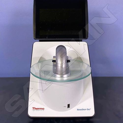
×
Add a mobile number to receive call from "Saaskin Corporation Private Limited."
×
Enter OTP
×
Share additional details for a quick response
×
Thank You!
Thank You for your valuable time. We have received your details and will get back to you shortly.
×
Get a price quote for Olympus BX43 System Microscope
×
- Saaskin Corporation Private Limited.
GST : 33AAZCS6003M1ZU
- 275/184, 1st Floor, Office No: 2, Golden Enclave, Periyar E V R Salai, P.H. Road, Kilpauk,Chennai - 600010, Tamil Nadu, India
- Mobile : 0091 9840819191 / 9940116677
- Phone:07971189171
- Mr Rajesh H (Business Operations Manager)
- Mobile:07971189171
|
-
 Accepts only Export inquiries
info@saaskin.com
Accepts only Export inquiries
info@saaskin.com
 |
SAASKIN CORPORATION PRIVATE LIMITED
All Rights Reserved.(Terms of Use) Developed and Managed by Infocom Network Private Limited. |
✕
Contact Us
Name
Comapny Name
Phone Number
Email Id
City / State

OTP Verification
Verification Code
Did not receive yet?
Resend OTP
Thank you!
We have received your requirements
For an immediate response, please call this
number 07971189171
✕
Tell us about your requirement
Price:
Quantity
Select Unit
Additional detail
Mobile number
Email
Name
Comapny Name
Phone Number
Email Id
City / State
Confirm Your Requirement
Verification Code
Did not receive yet?
Resend OTP
You’re Done!
We have received your requirements and will reply shortly with the best price.
Products You May Like